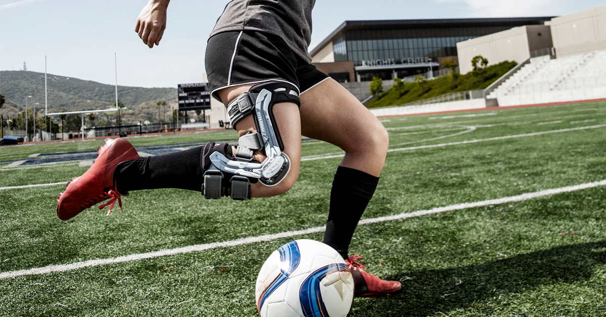
With the World Cup kicking off in Australia and New Zealand this year, the popularity of women’s football (soccer) is at an all-time high, with more women and girls getting involved every day. However, female football players are at a higher risk of anterior cruciate ligament (ACL) injuries compared to their male counterparts.1 Why are there more ACL injuries in women’s football, and how can knee bracing help in prevention and protection?
What is an ACL injury and how do they happen?
The anterior cruciate ligament (ACL) is one of the major stabilizing ligaments in the knee joint. Running diagonally in the middle of the knee, it prevents the tibia from sliding out in front of the femur and provides rotational stability to the knee.
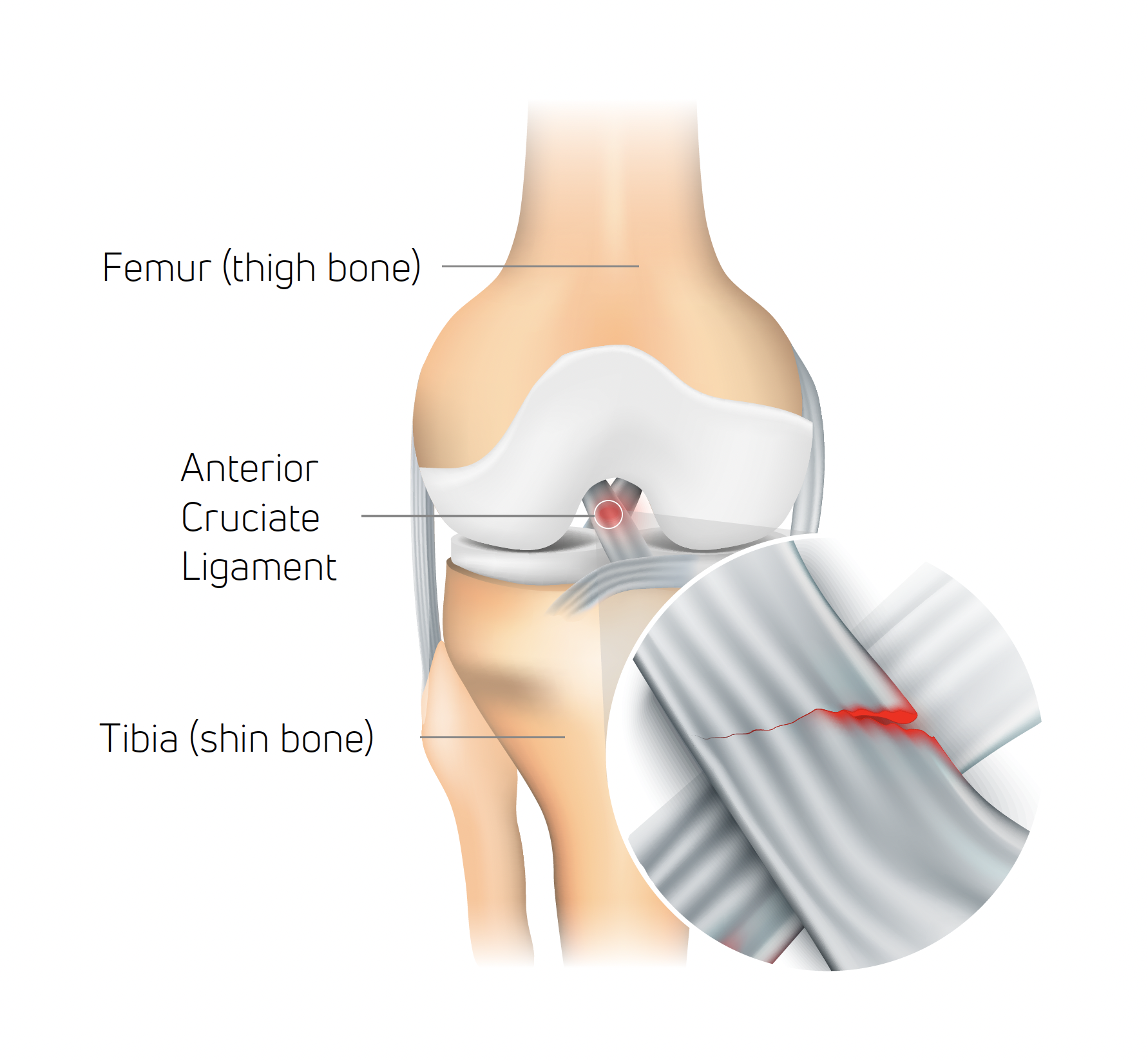
Although the ACL can be torn when the knee receives excessive external force, unlike most other ligaments it can also be injured in non-contact situations, with the latter accounting for 70% of all reported ACL injuries.2
In football and other field/court sports, non‐contact ACL injury typically involves a sharp stop, a sudden change of direction, or landing from a jump with inadequate knee and hip flexion (at or near full extension).1 The most common occurrence is from a combination of a deceleration manoeuvre with a change of direction while the foot is planted and the knee is nearly or fully extended; the excessive torsional force that results from the player trying to change direction can potentially strain or rupture the ACL.
Why ACL injuries are more common in female footballers
Studies have shown that female athletes are two- to ten-times more likely to suffer an ACL injury than male athletes.1 The phenomenon has had a noticeable impact on the 2023 Women’s World Cup, with several high-profile players missing out on the tournament as a result. There are a number of factors involved.
Differences in anatomy
The anatomy of female players predisposes them to an increased incidence of ACL injuries. Women generally have slimmer muscles, and a more considerable pelvic angle than men. The Q-angle, which refers to the angle formed between the hip and the knee, is higher in females, leading to more exertion placed on the ACL. The variance in the anatomy of the knee joint and surrounding bones results in lower overall knee stability, amplified by repetitive jumps or twists.
Hormonal predisposition
Research has suggested that the menstrual cycle poses a higher risk for ACL injuries during ovulation.3 Estrogen levels are higher during ovulation; high estrogen levels alter the collagen production, resulting in increased ligament laxity which can lead to decreased levels of overall knee stability.
Biomechanical Aspects
Studies have revealed distinct differences in landing techniques between men and women following a jump or leap.4 Females have landing mechanisms which increase the pressure placed on their knees. Furthermore, females are more likely than males to land with knee valgus position, making it harder to control the forces and positions during motions leading to tearing of the ACL.
How a knee brace can help reduce ACL injuries
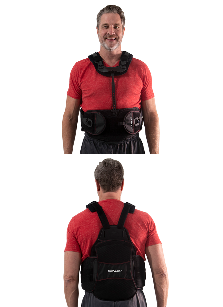
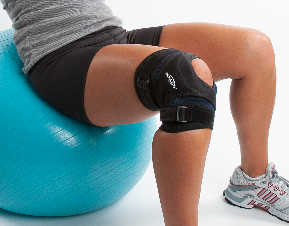
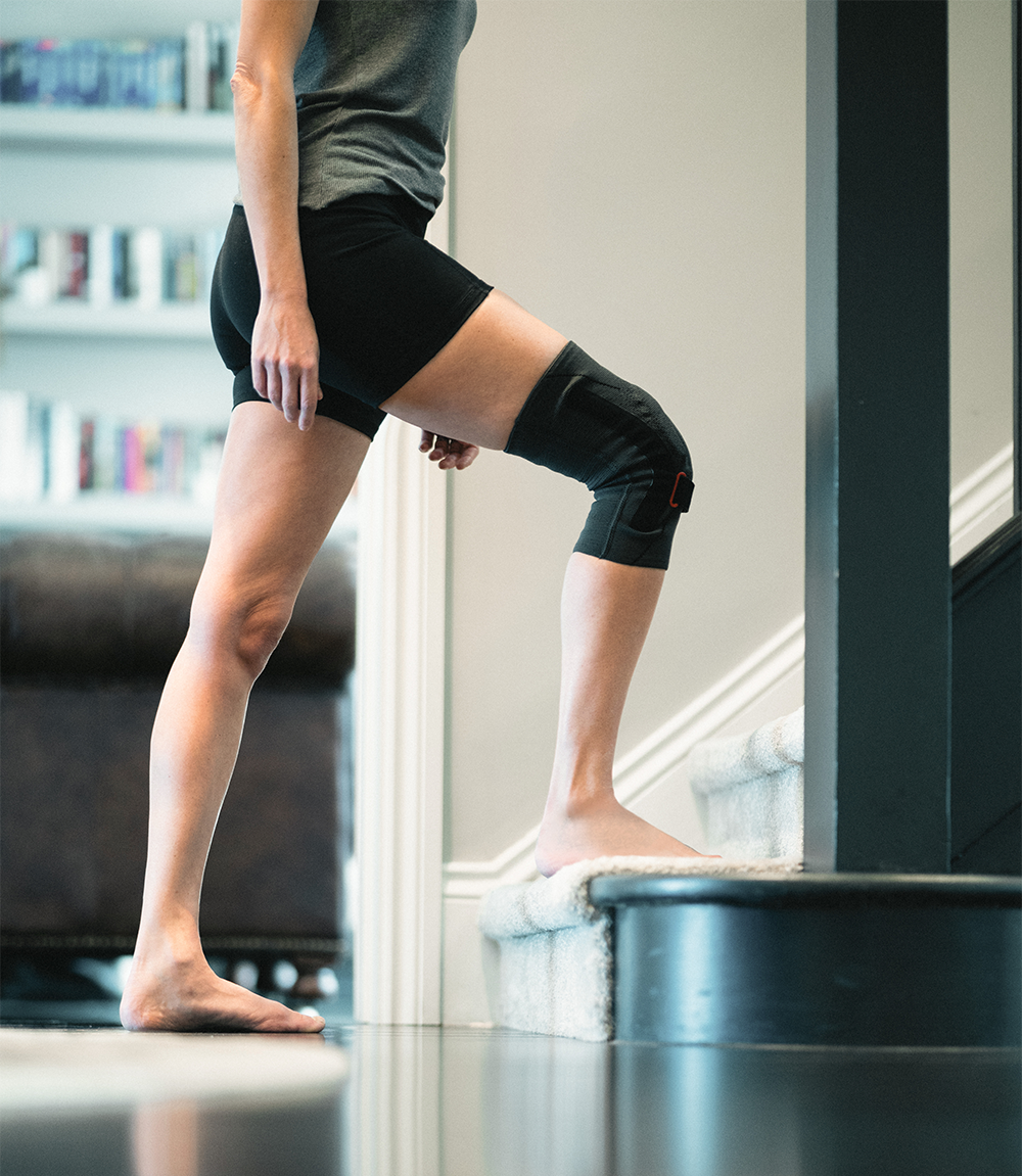
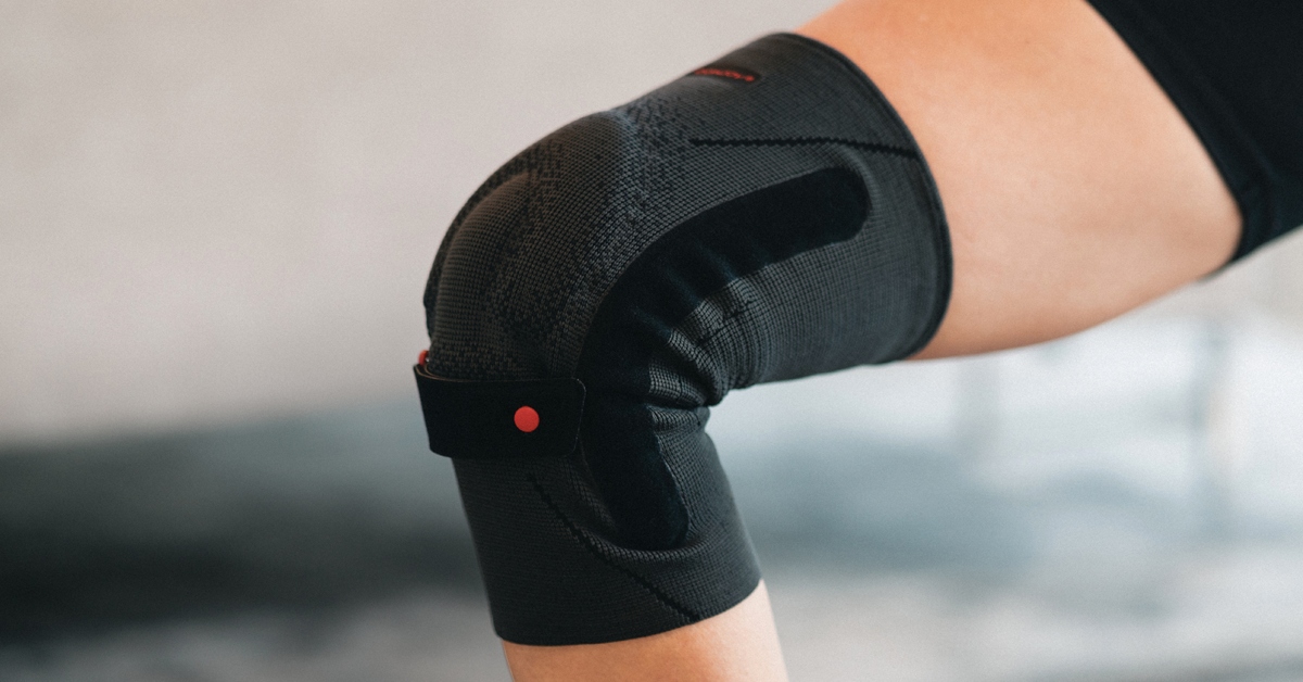
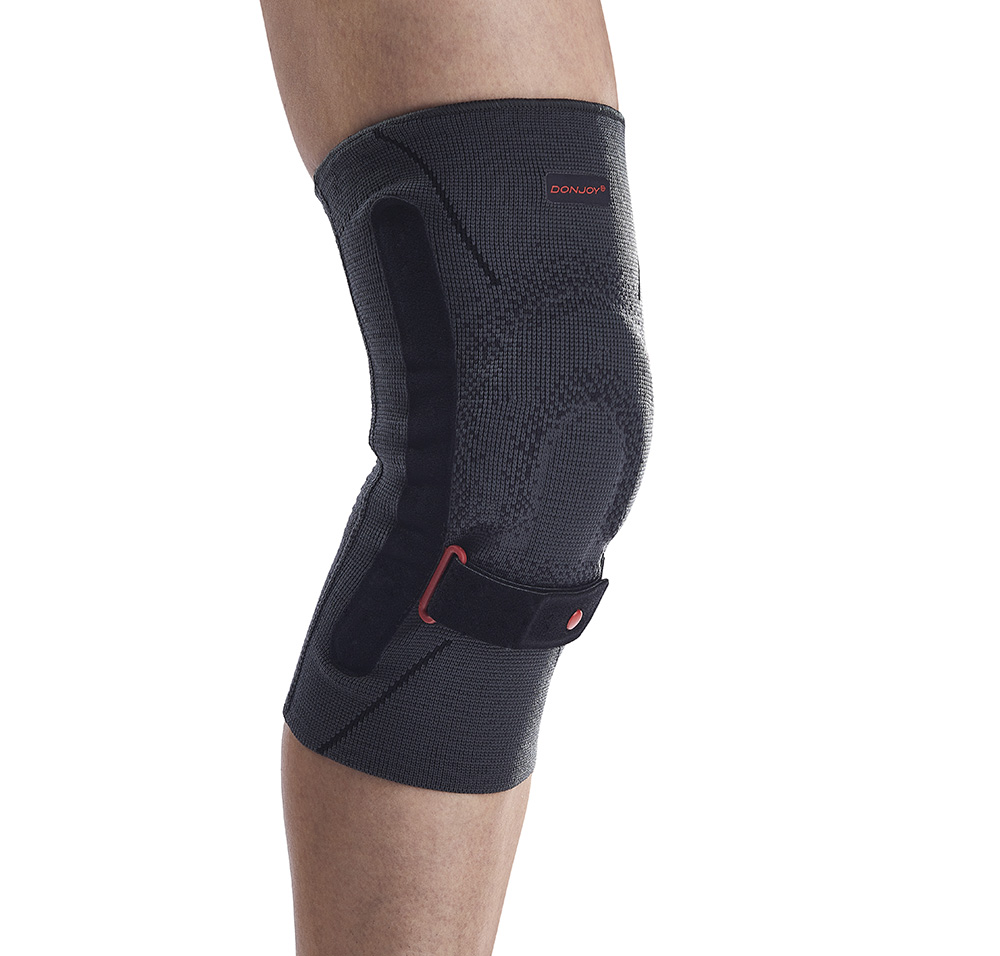
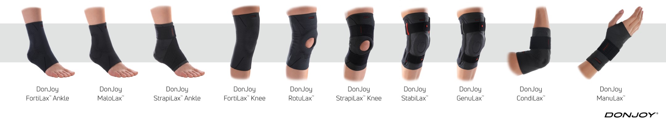
Clinical studies have demonstrated that wearing a knee brace during activity can help prevent ACL injury as well as protect against reinjury.5,6,7 DonJoy® knee braces utilize patented technology that reduces ACL strain.
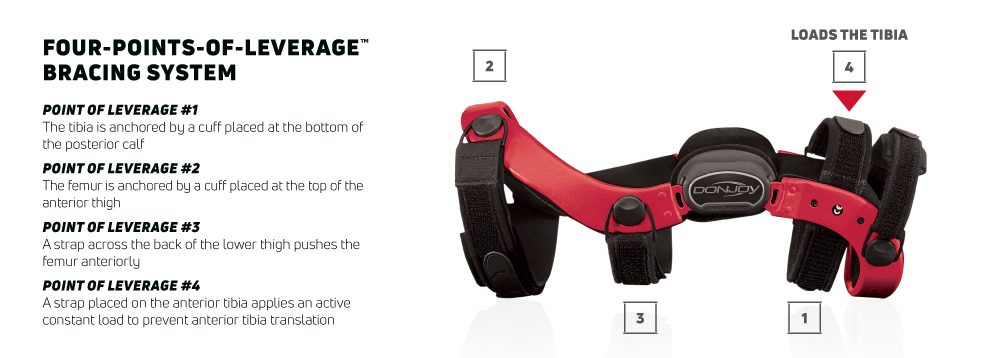
The Four-Points-of-Leverage™ system featured in DonJoy knee braces consists of a rigid cuff and strap configuration. Through this, a posterior force is applied to the tibia, which prevents anterior movement and reduces the strain on the ACL.8
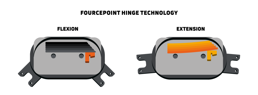
FourcePoint® hinge technology works to enhance DonJoy’s Four-Points-of-Leverage design by damping knee joint extension, which improves the mechanical performance of the brace and reduces shear forces at the knee. Addressing the “at-risk” position, the hinge resistance engages in the last 25 degrees of knee extension.
The FourcePoint hinge combined with the Four-Points-of-Leverage cuff and strapping design provides a more comfortable brace that reduces anterior shear forces at the knee. The stability this provides is beneficial for both female football players looking to avoid ACL injuries during training, and those who are recovering from an existing injury, as it reduces strain on the healing ACL graft.9,10
To find out more about DonJoy knee braces, go to enovis-medtech.eu.
References
- Silvers, H. J., & Mandelbaum, B. R. (2007). Prevention of anterior cruciate ligament injury in the female athlete. British journal of sports medicine, 41 Suppl 1(Suppl 1), i52–i59.
- Arendt EA, Agel J, Dick R. Anterior cruciate ligament injury patterns among collegiate men and women. J Athl Train 1999;34(2):86-92.
- Yu, W. D., Liu, S. H., Hatch, J. D., Panossian, V., & Finerman, G. A. (1999). Effect of estrogen on cellular metabolism of the human anterior cruciate ligament. Clinical orthopaedics and related research, (366), 229–238.
- Butler, R. J., Willson, J. D., Fowler, D., & Queen, R. M. (2013). Gender differences in landing mechanics vary depending on the type of landing. Clinical journal of sport medicine : official journal of the Canadian Academy of Sport Medicine, 23(1), 52–57.
- Tuang, B.H.H., Ng, Z.Q., Li, J.Z., Sirisena D. (2023). Biomechanical Effects of Prophylactic Knee Bracing on Anterior Cruciate Ligament Injury Risk: A Systematic Review. Clin J Sport Med. Jan 1;33(1):78-89.
- Perrone, G.S., Webster, K.E., Imbriaco, C., Portilla, G.M., Vairagade, A., Murray, M.M., Kiapour, A.M. (2019). Risk of Secondary ACL Injury in Adolescents Prescribed Functional Bracing After ACL Reconstruction. Orthop J Sports Med. Nov 12;7(11):2325967119879880.
- Bodendorfer, B.M., Anoushiravani, A.A., Feeley, B.T., Gallo, R.A. (2013). Anterior cruciate ligament bracing: evidence in providing stability and preventing injury or graft re-rupture. Phys Sportsmed. Sep;41(3):92-102.
- Fleming, B. C., Renstrom, P. A., Beynnon, B. D., Engstrom, B., & Peura, G. (2000). The influence of functional knee bracing on the anterior cruciate ligament strain biomechanics in weightbearing and nonweightbearing knees. The American journal of sports medicine, 28(6), 815–824.
- Théoret, D., & Lamontagne, M. (2006). Study on three-dimensional kinematics and electromyography of ACL deficient knee participants wearing a functional knee brace during running. Knee surgery, sports traumatology, arthroscopy : official journal of the ESSKA, 14(6), 555–563.
- Stanley, C. J., Creighton, R. A., Gross, M. T., Garrett, W. E., & Yu, B. (2011). Effects of a knee extension constraint brace on lower extremity movements after ACL reconstruction. Clinical orthopaedics and related research, 469(6), 1774–1780.