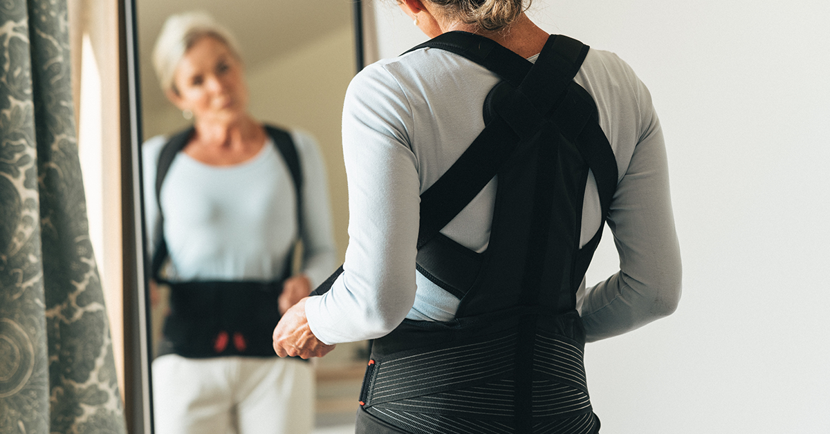
Knee osteoarthritis (OA) is one of the most widespread musculoskeletal conditions in the world — and its impact on daily life can be profound. Whether you are a patient struggling to walk to the local shops, or a clinician searching for effective conservative management options, understanding the role of knee OA bracing is increasingly important. The evidence is compelling: the right brace, fitted correctly, can reduce pain, improve mobility, and in some cases delay or even prevent the need for surgery.
Knee OA: A Background
Knee OA affects an estimated 22.9% of people aged 40 and over — that is 654 million people globally in this age group alone.1 Radiographic evidence of knee OA is even more prevalent, detected in 28.7% of the population across all ages, though not all cases are symptomatic.1 The World Health Organization estimates that 344 million people living with osteoarthritis (across all joints) experience moderate or severe levels that could benefit from rehabilitation.2
For many patients, the journey from early symptoms to the operating theatre is a long one. Research by London et al. calculated that approximately 20% of American patients with symptomatic knee OA linger in what is known as the “treatment gap” — the period after conservative treatments have been exhausted but before surgical intervention — for up to 10 years. For younger patients, this gap can stretch to 20 years, a significant period of pain, reduced activity, and diminishing quality of life.3
Understanding the Vicious Cycle of Knee OA
Knee OA does not progress in a straight line. It is driven by a self-reinforcing cycle of interconnected problems. Malalignment of the knee — where the joint is not tracking correctly — leads to aberrant biomechanics and increased compartment loading, which in turn causes pain. Pain leads to decreased activity, which contributes to weight gain and muscle weakness, which further destabilizes the joint, and so the cycle continues.
Conventional pharmacological approaches such as analgesics and Non-Steroidal Anti-Inflammatory Drugs (NSAIDs) can reduce pain and improve quality of life, but they do not address the underlying biomechanical causes of OA. There is also evidence that increased pain-free activity or walking speed following medication use may actually lead to increased joint loading4 and accelerated disease progression5 — a counterintuitive but important consideration for clinicians.
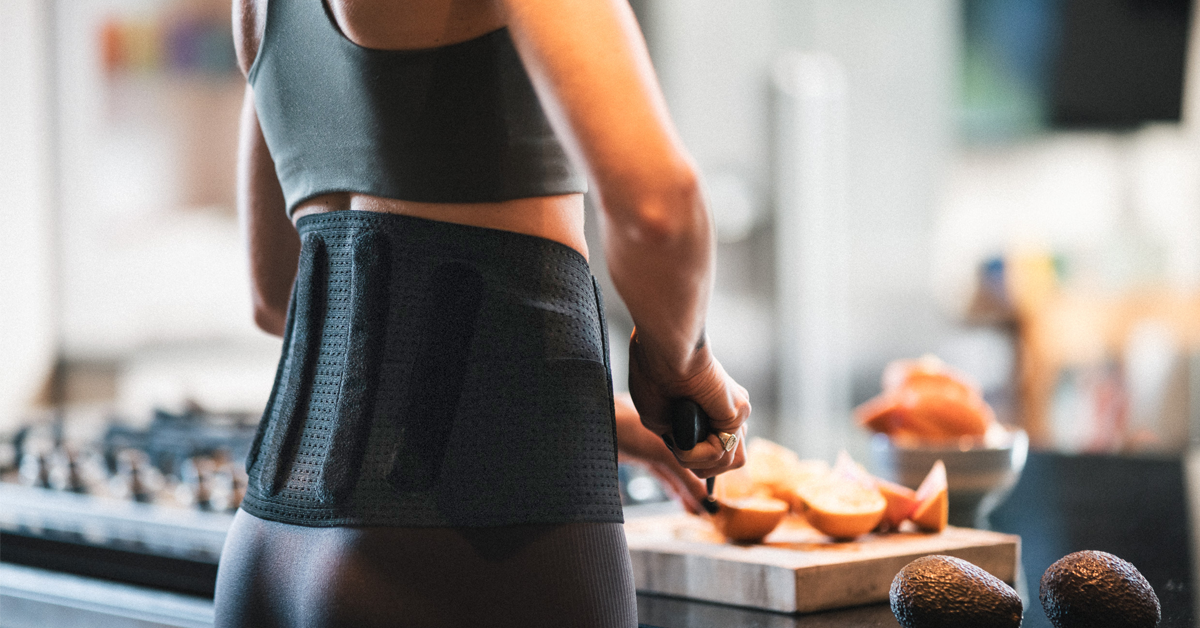
This is where off-loading knee bracing offers something fundamentally different.
How Knee OA Bracing Can Break the Cycle
A well-designed knee OA brace works by physically altering the forces acting on the knee during movement. The clinical evidence supporting this mechanism is substantial.
Increasing joint space
Research using highly accurate biplane radiography demonstrated in patients with medial knee OA that wearing a DonJoy OA Defiance brace can induce a significant increase of 0.3 mm in medial compartment dynamic joint space during gait — roughly a 10% increase during the impact phase of walking. This improvement was consistent from heel strike to terminal stance, meaning the joint is protected throughout the most demanding part of each step.6
Correcting malalignment
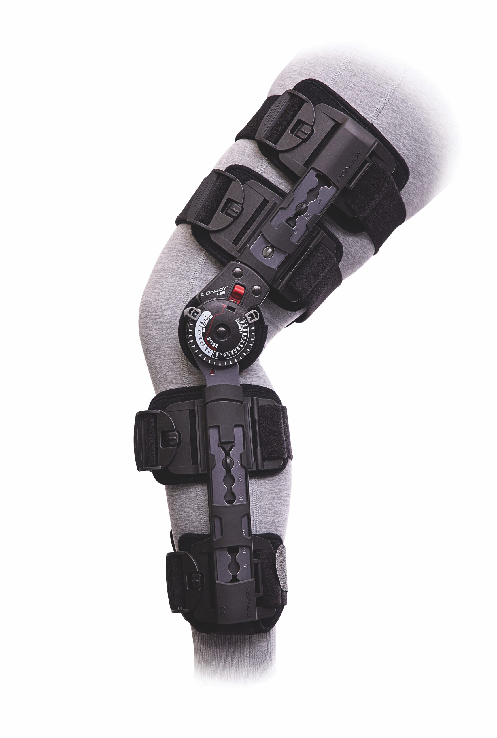
OA braces have been shown to shift the knee into a more corrected position in patients with varus (bow-legged) knees7,8 with the greatest corrective effect occurring at approximately 10% of the gait cycle — precisely the point of greatest loading.8,9 A biomechanical study using the OA Adjuster brace found a difference of 4° in varus angle between braced and unbraced conditions, a clinically meaningful correction.9
Reducing knee adduction moment
The knee adduction moment — the force that drives compression on the medial side of the joint — is a key driver of medial compartment OA progression. OA braces have been shown to reduce peak knee adduction moment by up to 32% during gait,7,9,10 with greater reductions achieved when the brace hinge is dialled in further.10 The adduction impulse, which accounts for both load and loading time, can be reduced by up to 37%.10
Improving stability and confidence
Muscle weakness and decreased stability contribute to reduced patient confidence in movement. OA bracing has been shown to improve both perceived and objective knee stability,11,12,13 which encourages patients to move more — breaking the inactivity cycle rather than perpetuating it.
The Impact of Knee OA on Pain and Everyday Life
The clinical outcomes for pain relief are striking. In a systematic review by Feehan et al. that included 15 clinical studies, 98.6% of 567 patients with medial knee OA experienced pain relief when fitted with an off-loading brace.14 In another study, both custom and off-the-shelf brace options produced significant reductions in pain and stiffness, with custom bracing also showing meaningful improvements in function.7
The effect on daily mobility is equally significant. A patient feedback study by Dries et al. (2022) found that wearing an OA Defiance brace considerably expanded mobility across all patient groups.15 The proportion of patients confined to their home environment reduced by 74%, while 42% of brace wearers reported being able to take a long walk or visit a local shop — activities that had previously been out of reach. This is not a minor quality-of-life improvement; for many patients, it represents a return to independence.
Can Knee OA Bracing Delay Surgery?
One of the most important questions for both patients and healthcare systems is whether bracing can delay or reduce the need for surgical intervention. The evidence here is encouraging.
A study by Lee et al. (2017) found that patients who wore an off-loading knee brace for two years or more did not require surgery at eight-year follow-up.16 Given that the off-loading knee brace is a cost-effective treatment option, its potential as a bridging therapy — reducing the burden of the treatment gap on both patients and healthcare systems — is considerable.16,17
A Place in Every Stage of Treatment
Modern OA bracing is not a one-size-fits-all solution. Braces are now designed to address a spectrum of OA severity, from early-stage support through to moderate and severe disease. This means clinicians can tailor brace selection to the individual patient’s needs, activity level, and degree of joint involvement.
For patients, the message is equally clear: living with knee OA does not have to mean accepting a steady decline in mobility and independence. For healthcare professionals, the evidence supports placing OA bracing earlier and more consistently in the conservative management pathway — not as a last resort before surgery, but as an active intervention that can protect the joint, relieve pain, and keep patients moving.
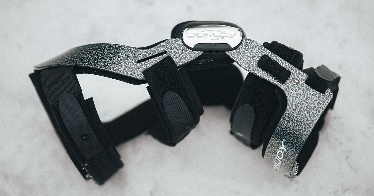
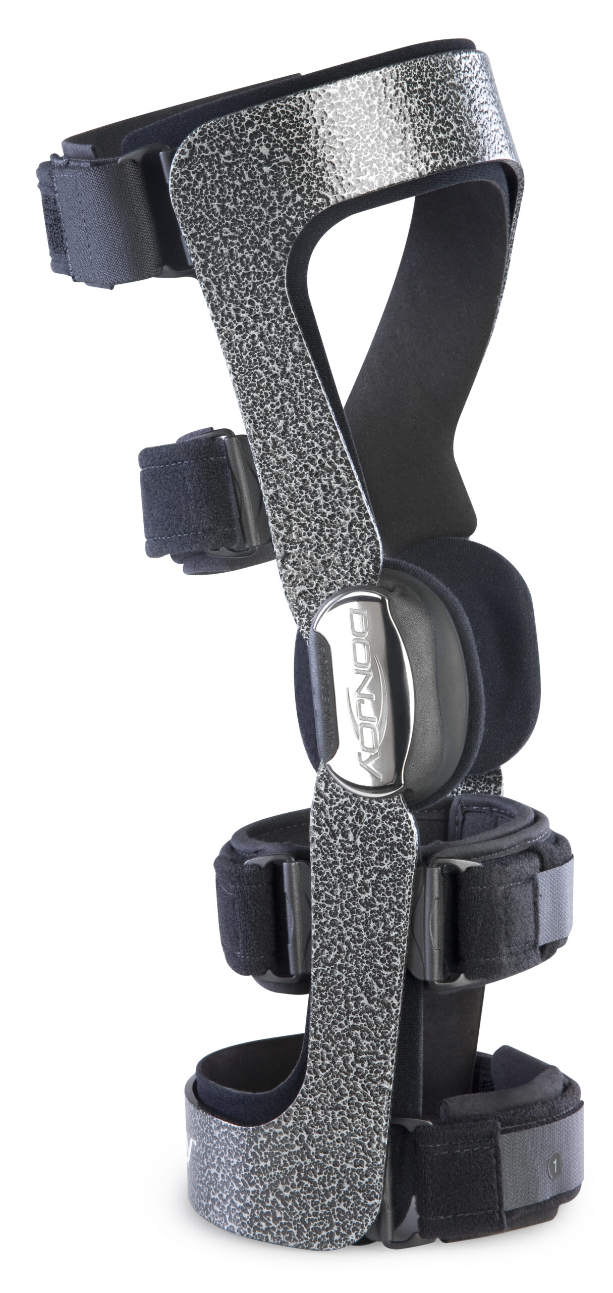
DonJoy® Knee OA Braces: For Every Stage of Osteoarthritis
Under the DonJoy® brand, Enovis™ offers a full portfolio of braces for all severities of knee OA and levels of patient activity.
DonJoy OA GO® helps provide relief from pain and mobility issues caused by mild to moderate knee osteoarthritis.17 With a simple twist of a dial, this innovative soft knee brace’s three-point offloading system quickly offloads the affected compartment to help relieve pain and ease movement.
ROAM™ OA helps patients say goodbye to knee pain. From picking up their grandkids to teeing off at the driving range, lightweight, low-profile joint offloading and support never felt so good—or was so easy to prescribe. ROAM helps improve mobility and provides relief by offloading the pressure of unicompartmental osteoarthritis or other knee pain.17
OA Nano™ is our lightest functional knee brace for mild to moderate osteoarthritis. Powered by DonJoy’s clinically proven Adjuster™ technology,17 it combines targeted offloading with minimal weight, helping patients maintain their activity levels in comfort. The flexibility of the streamlined aluminum frame allows for an intimate fit while providing offloading to support pain relief. OA Nano is designed to help people stay active and move freely, making patient compliance a reality.
For more information about managing knee osteoarthritis and finding the right support, visit donjoyoabraces.com.
Healthcare professionals interested in learning more about knee OA braces can contact their local Enovis representative or visit our website for detailed product information.
References
- Cui A, Li H, Wang D, et al. Global, regional prevalence, incidence and risk factors of knee osteoarthritis in population-based studies. EClinicalMedicine. 2020;29–30:100587.
- WHO July 2023 Osteoarthritis Key Facts. https://www.who.int/news-room/fact-sheets/detail/osteoarthritis
- London NJ, Miller LE, Block JE. Clinical and economic consequences of the treatment gap in knee osteoarthritis management. Med Hypotheses. 2011;76(6):887–92.
- Schnitzer TJ, Popovich JM, Anderson GBJ, Andriacchi TP. Effect of piroxicam on gait in patients with osteoarthritis of the knee. Arthritis and Rheumatism. 1993;9:1207–13.
- Huskisson EC, Berry H, Gishen P, et al. Effects of antiinflammatory drugs on the progression of osteoarthritis of the knee. Journal of Rheumatology. 1995;22:1941–6.
- Nagai K, Yang S, Fu FH, Anderst W. Unloader knee brace increases medial compartment joint space during gait in knee osteoarthritis patients. Knee Surg Sports Traumatol Arthrosc. 2019;27(7):2354–2360.
- Draganich L, Reider B, Rimington T, et al. The effectiveness of self-adjustable custom and off-the-shelf bracing in the treatment of varus gonarthrosis. J Bone Joint Surg Am. 2006;88(12):2645–52.
- Richards J, Jones R, Kim W. Biomechanical changes in the conservative treatment of medial compartment osteoarthritis of the knee using valgus bracing. ICRS 2006.
- The Comprehensive Textbook of Clinical Biomechanics, 2nd Edition. Elsevier 2018.
- Orishimo KF, Kremenic IJ, Lee SJ, et al. Is valgus unloader bracing effective in normally aligned individuals. Knee Surg Sports Traumatol Arthrosc. 2013;21(12):2661–6.
- Hart HF, Collins NJ, Ackland DC, et al. Immediate Effects of a Brace on Gait Biomechanics for Predominant Lateral Knee Osteoarthritis and Valgus Malalignment After ACL Reconstruction. Am J Sports Med. 2016;44(4):865–73.
- Hart HF, Crossley KM, Collins NJ, Ackland DC. Bracing of the Reconstructed and Osteoarthritic Knee during High Dynamic Load Tasks. Med Sci Sports Exerc. 2017;49(6):1086–1096.
- Kwaees TA, Richards J, Rawlinson G, et al. Can the use of proprioceptive knee braces have implications in the management of osteoarthritic knees. Prosthet Orthot Int. 2019;43(2):140–147.
- Feehan NL, Trexler GS, Barringer WJ. The Effectiveness of Off-Loading Knee Orthoses in the Reduction of Pain in Medial Compartment Knee Osteoarthritis: A Systematic Review. J Prosthet Orthot. 2012;24(1):39–49.
- Dries T, Van Der Windt JW, Akkerman W, et al. Effects of a semi-rigid knee brace on mobility and pain in people with knee osteoarthritis. J Rehabil Med Clin Commun. 2022;5:2483.
- Lee PY, Winfield TG, Harris SR, et al. Unloading knee brace is a cost-effective method to bridge and delay surgery in unicompartmental knee arthritis. BMJ Open Sport Exerc Med. 2017;2(1):e000195.
- Mistry DA, Chandratreya A, Lee PYF. An Update on Unloading Knee Braces in the Treatment of Unicompartmental Knee Osteoarthritis from the Last 10 Years: A Literature Review. Surg J (N Y). 2018;4(3):e110–e118.