Achilles tendonitis, also known as Achilles tendinopathy, is an overuse injury affecting the Achilles tendon. It is common among athletes and those engaged in repetitive physical activity, such as running and jumping. This article explains the causes and symptoms of Achilles tendonitis and explores the available treatments.
Causes
Achilles tendonitis often arises from excessive strain on the tendon due to overuse, improper footwear, or poor biomechanics (such as flat feet). Other contributing factors include sudden increases in activity level, weak calf muscles, or tightness in the calf or hamstring.
Athletes who engage in sports like running, basketball, or football are particularly prone to this injury, though it can also occur in people who do not warm up properly or who have abnormal foot posture.
Symptoms
The symptoms of Achilles tendonitis include pain, tenderness, and stiffness in the tendon, particularly at the back of the heel. Pain is often worse in the morning or after periods of inactivity and can be made worse by physical activity.
In some cases, swelling or thickening around the tendon may be visible, and in more severe cases, nodules may form along the tendon.
Prevention, treatment, and management of Achilles tendonitis
Preventing Achilles tendonitis involves ensuring proper training techniques, including gradual increases in the intensity and amount of exercise. Wearing appropriate footwear, stretching before physical activity, and strengthening the calf muscles can also reduce the risk of injury.
If Achilles tendonitis is already present, treatment is the next step. This generally starts with conservative measures like rest, ice, and anti-inflammatory medications. Physical therapy is crucial in managing Achilles tendonitis, particularly eccentric strengthening exercises that target the calf muscles to reduce strain on the Achilles tendon. Other interventions may include orthotics such as braces or heel lifts to reduce tendon strain, and taping to support the area during activity.
In more persistent cases, advanced treatments like shock wave therapy may be considered. Surgical intervention is typically a last resort for chronic, unresponsive cases.
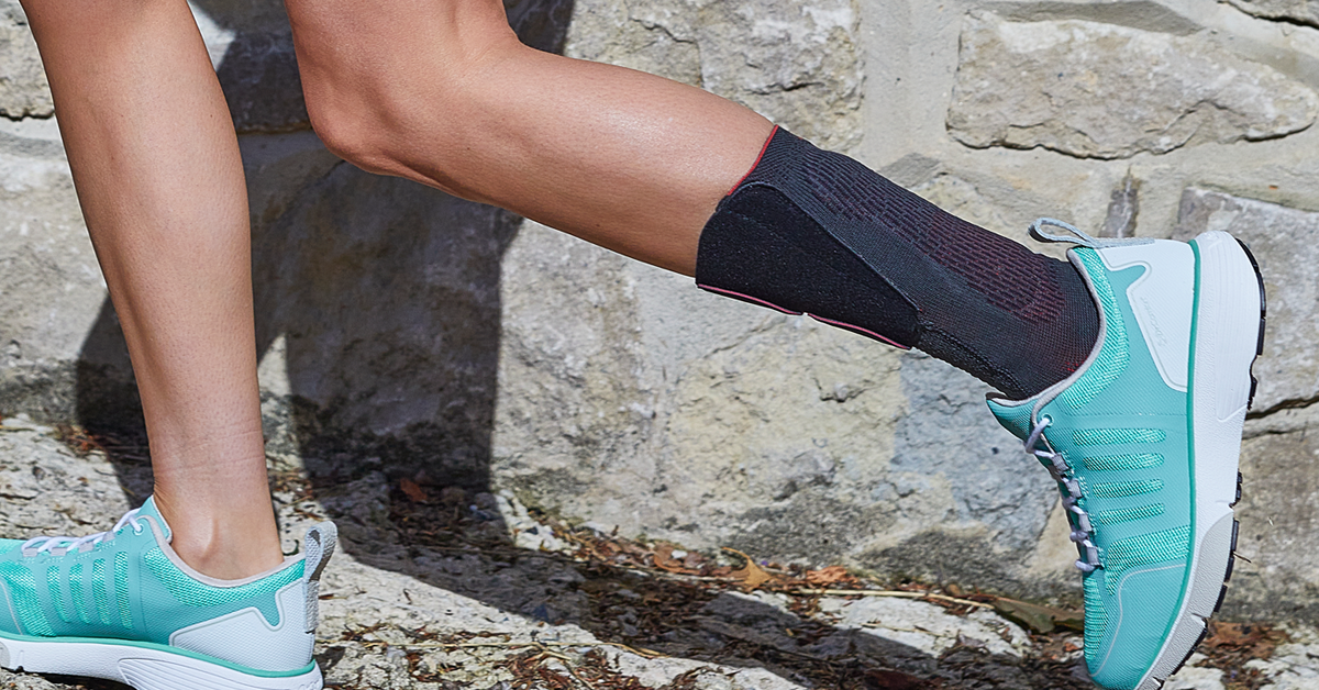
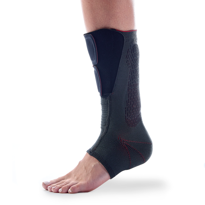
Introducing AchilloForce® by DonJoy®: a new ankle support for Achilles tendonitis
For patients suffering with Achilles tendonitis, a new product by DonJoy® may offer relief. AchilloForce® is an elastic knitted ankle brace with an integrated pad covering the Achilles tendon area. This combination of compressive sleeve and cushioning pad can help deliver comfortable and easy relief and proprioception for Achilles tendon pain and inflammation.
The brace simply slides over the foot (with or without a sock on underneath) and is fixed in place by a simple two-part hook-and-loop closure at the front. Because of its slim profile, it fits easily into a shoe, and its breathable materials help the foot stay cool and dry during wear.
What’s more, AchilloForce comes with a pair of silicone heel wedges – placing one of these into the shoe underneath the heel provides extra cushioning for further relief of the Achilles tendon.
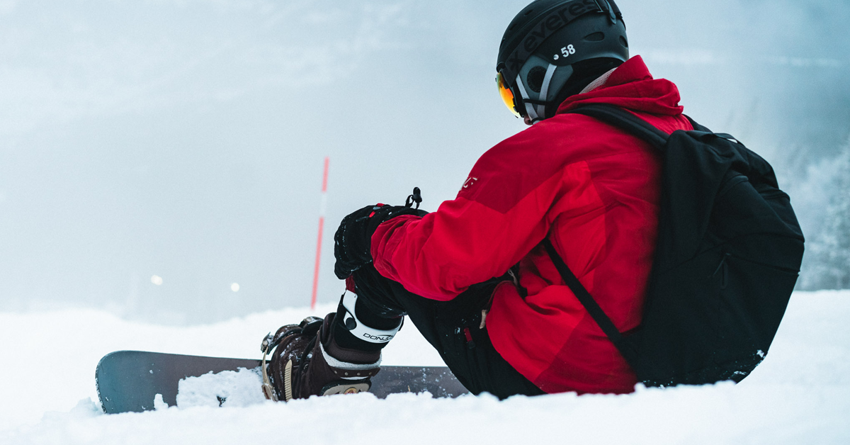
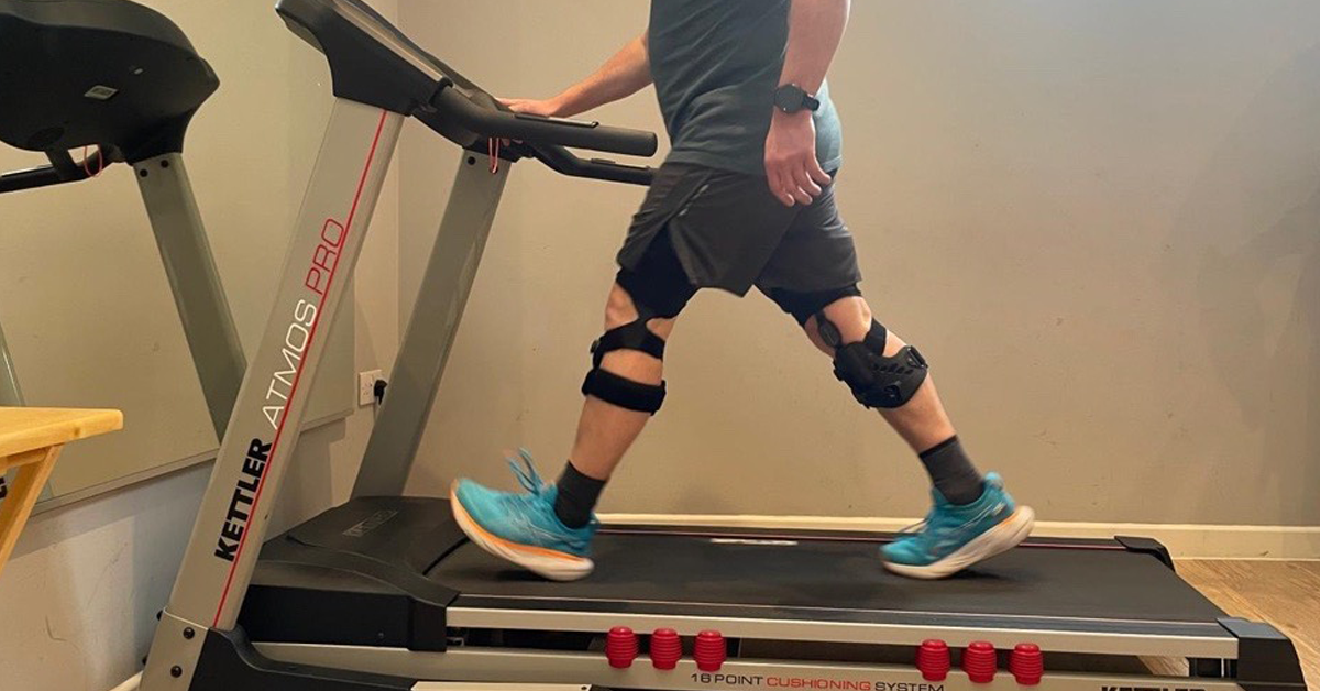
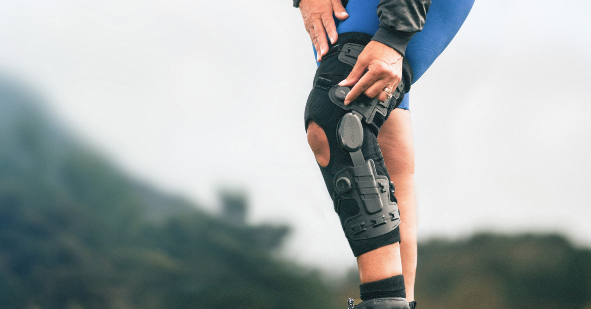
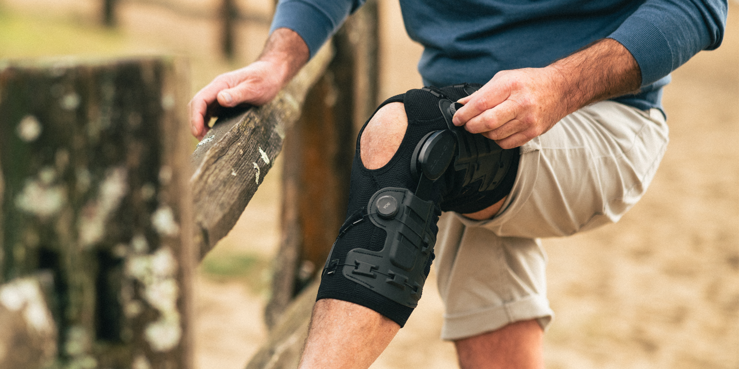
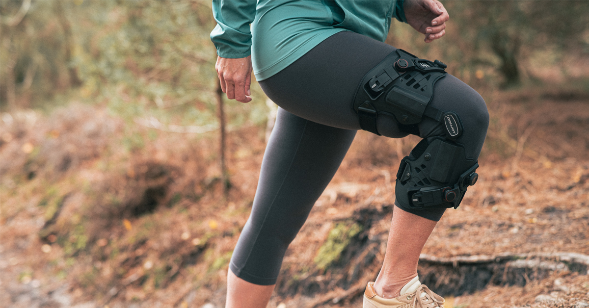
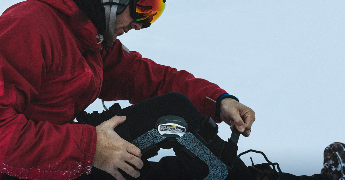
With winter around the corner, skiers and snowboarders are gearing up for a fresh season on the slopes. These snow sports provide a unique mix of speed, agility, and excitement—but they also bring a high risk of knee injuries, especially with the sharp turns, jumps, and high-impact landings. Whether you’re tackling moguls on skis or carving down a snowboard terrain park, the Defiance® PRO knee brace from DonJoy® offers robust protection, stability, and injury prevention for snow enthusiasts of all kinds.
Why skiers and snowboarders need knee support
Knee injuries are among the most common injuries for both skiers and snowboarders1. The rapid twisting motions in skiing, coupled with the board-fixed position in snowboarding, can put intense stress on the knee joint, especially the anterior cruciate ligament (ACL) and medial collateral ligament (MCL). Add high speeds, dynamic movement, and unpredictable falls, and it’s easy to see why additional knee protection is essential. The Defiance PRO knee brace is designed to provide that vital support and stability, whether you’re carving powder on a snowboard or navigating steep ski trails.
Key features of the Defiance® PRO for snow sports
The Defiance PRO knee brace combines clinically proven technology with a lightweight build to provide stability, prevent injury, and enhance performance without limiting your range of motion. Here’s a closer look at how this knee brace supports skiers and snowboarders alike:
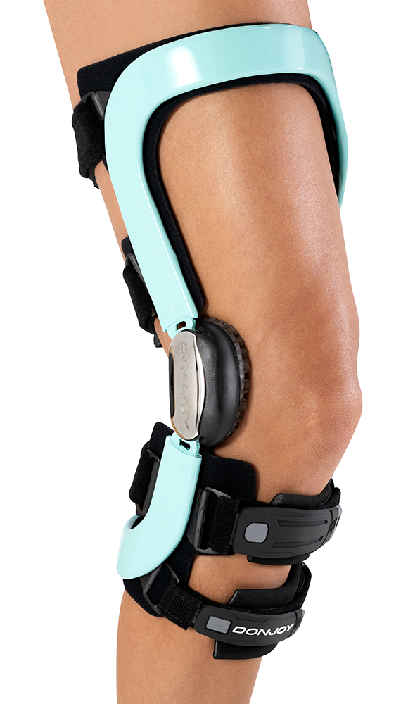
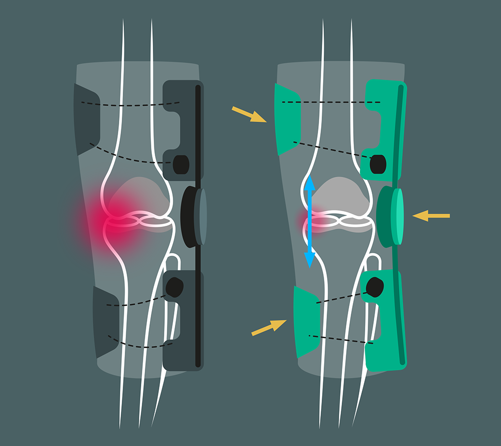
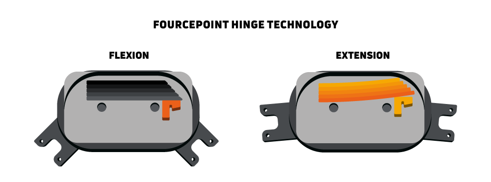
1. FourcePoint® hinge technology
The brace’s FourcePoint® hinge technology is clinically proven to help protect the knee from injury2. By keeping the knee out of full and hyperextension, its dampening mechanism helps reduce the strain on the ACL that can occur during the impacts of skiing and snowboarding2.
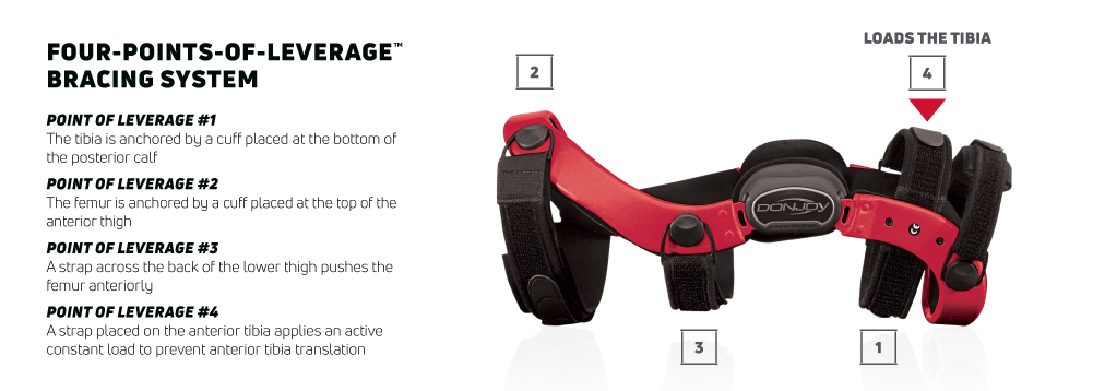
2. Four-Points-of-Leverage™ system
The ACL is a ligament particularly vulnerable in skiing and snowboarding. The Defiance PRO’s patented Four-Points-of-Leverage™ system is clinically proven to reduce strain on the ACL by helping to prevent forward shin movement.3 For snowboarders, who face different movement patterns but similar knee stresses, this system provides the same stability against unanticipated jerks or impacts.
3. Custom fit for comfort and stability
As well as utilizing anti-migration technology, every Defiance PRO is custom-made to fit each user’s leg shape. This is crucial for skiers and snowboarders who need a stable fit without sliding or shifting. For both sports, this stability can make all the difference on sharp turns, landings, or during intense runs.
4. Lightweight carbon fiber frame
Made from carbon fiber, the Defiance PRO is engineered to withstand high impact while remaining lightweight and low-profile. Skiers and snowboarders can wear it comfortably under their snow gear without feeling weighed down, making it ideal for long sessions on the slopes. The durable frame offers protection even in rough conditions, providing peace of mind for those aiming to push their limits.
What’s new about Defiance PRO?
Skiers and snowboarders who have worn knee braces in the past may already be familiar with the name of Defiance. Building on its legacy, the new Defiance PRO retains all the features users have come to know, while introducing several enhancements.
Even lower profile
Compared to the Defiance Classic, the profile of Defiance PRO is slimmer by 5 mm, making for an even more discrete fit.
Internally mounted swiveling straps
On the new Defiance PRO, the straps are attached to the inside of the frame for a cleaner profile with less possible friction. The straps can also swivel at these connection points, helping to provide a degree of dynamic movement while retaining that all-important stability. And with their new soft-touch ends, the straps are even easier to apply and keep fastened.
More comfortable condyle pads
Defiance PRO features new condyle pads made from soft silicone for a more comfortable contact point with the knee.
New improved liners
The Defiance PRO’s new C-6 liner material is soft to the touch, moisture wicking, and anti-microbial, all of which helps keep wearers comfortable during use.
Preventing injury and supporting recovery
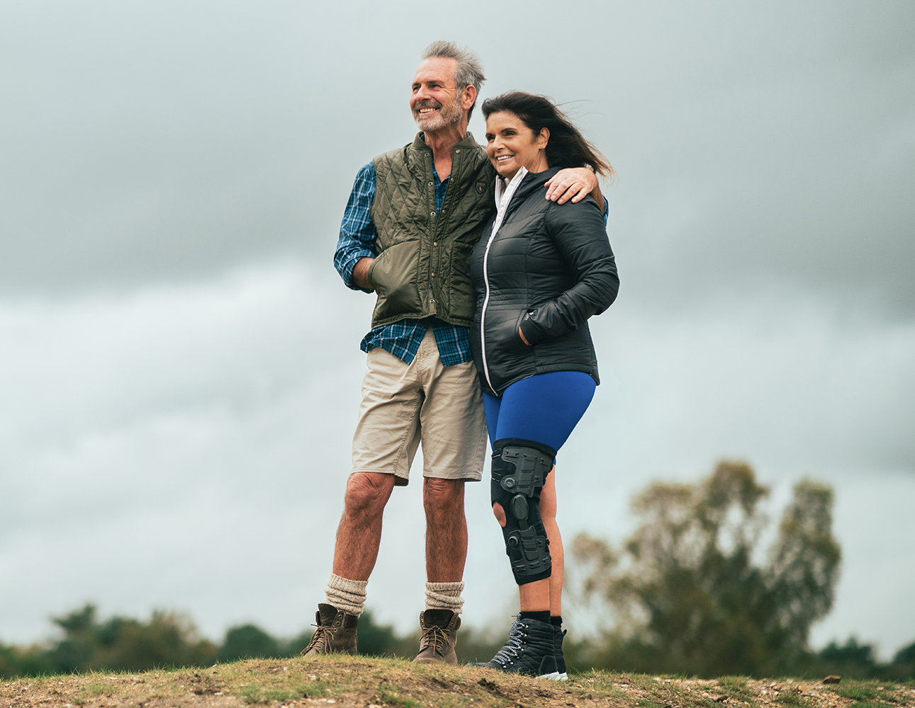
For skiers and snowboarders alike, injury prevention is vital to enjoying a long and active snow season. The Defiance PRO is crafted to help prevent both acute injuries (like ACL and MCL tears) and wear-and-tear injuries that can develop over time. Skiers, who face high-speed turns and quick directional changes, and snowboarders, who need support against hard impacts and rotational movements, can both benefit from the brace’s stabilizing features.
The Defiance PRO is also a helpful tool for those in recovery. If you’re returning to skiing or snowboarding after a knee injury, the brace’s targeted support can give you confidence to get back on the mountain. A re-injury rate of 5-10% for ACL injuries shows the importance of added protection4.
But don’t be fooled that this is something that only affects more mature people; secondary ACL injuries are common in adolescents too. However, at least one clinical study with young people has shown that wearing a knee brace can help prevent injury to ACL grafts following surgery.5
Make the most of your ski season with the Defiance PRO knee brace
As you gear up for ski and snowboard season, consider the added support and injury prevention that the Defiance PRO knee brace can offer. Whether you’re an experienced skier or a snowboarder hitting the terrain park, investing in a Defiance PRO can provide the security to allow you to take on the mountain without hesitation. Make this season one to remember, with knees that are fully supported for every twist, turn, and landing.
Wagner M et al. Incidence of alpine skiing and snowboarding injuries. Injury. 2023 Aug;54(8):110830.
Yu B et al. Immediate effects of a knee brace with a constraint to knee extension on knee kinematics and ground reaction forces in a stop-jump task. Am J Sports Med 2004;32(5):1136-43.
Fleming BC et al. The influence of functional knee bracing on the anterior cruciate ligament strain biomechanics in weightbearing and nonweightbearing knees. Am J Sports Med 2000;28(6):815-24.
Arendt EA et al. Anterior cruciate ligament injury patterns among collegiate men and women. Journal of Athletic Training. 1999;34(2):86-92.
Perrone GS et al. Risk of Secondary ACL Injury in Adolescents Prescribed Functional Bracing After ACL Reconstruction. Orthop J Sports Med. 2019;7(11):2325967119879880.
For orthotist Laura Aggett, delivering the best outcomes for her patients is more than just a priority—it’s a passion. As the owner and director of Lewis Brand Orthotics, a leading private orthotics practice with multiple locations in the UK, Laura constantly seeks innovative solutions that can enhance her patients’ quality of life. Her dedication frequently leads her to explore the latest advancements in orthopedic products, always aiming to find the best fit for her patients’ needs.
One of Laura’s most exciting recent discoveries is DonJoy’s new ROAM OA knee brace. Designed for individuals with moderate to severe knee osteoarthritis (OA), the ROAM OA brace offers both comfort and offloading to help alleviate pain. After learning about the product from Enovis bracing specialists, Laura was eager to offer it to her patients dealing with knee OA pain.
‘I’ve been really impressed with how the ROAM OA works and how effective it is for knee osteoarthritis patients,’ says Laura.
‘I’ve only received positive feedback from my patients [on ROAM OA].’
Laura Aggett, Lewis Brand Orthotics
A standout feature of the ROAM OA knee brace is its telescoping frame, which can be adjusted to accommodate patients with unique leg shapes or sizes. ‘Some patients find that certain braces rub uncomfortably on their thigh or don’t provide the necessary force to offload the affected knee compartment,’ Laura says. ‘With the ROAM OA’s telescoping frame, I can lengthen or shorten both the proximal and distal segments of the brace to ensure a better fit, or to spread the force over a greater area.’
Ease of use is at the heart of ROAM OA’s design philosophy. Given that knee OA primarily affects older adults who may also have dexterity issues1, it is essential that products tailored for them are straightforward to apply and adjust.
‘They’ve put a lot of thought into the design of the straps,’ says Laura. ‘The magnetic touch-close fastenings make it really easy for the patients. And my favorite part is the strap that leaves the knee crease free – this definitely improves comfort and compliance.’
The ROAM OA brace also features a combination of strategically placed straps and adjustable BOA dials. These work together with the unique condyle harness to create a pulling force on the knee to help offload the affected compartment. ‘You don’t have to unload it completely like some braces,’ Laura says. ‘Instead, you can both increase and decrease the load through the BOA system on a step-by-step basis. That gives patients a lot more adjustability and flexibility for different activities like going up and down hills.’
Having only recently launched, the full impact of the ROAM OA brace on knee osteoarthritis patients is yet to be seen. However, early adopters like Laura are optimistic about its potential. ‘So far, I’ve only received positive feedback from my patients,’ she says. ‘I’m looking forward to fitting many more in the years to come.’
Laura recently presented her experiences of knee bracing on a webinar with Enovis. You can watch the recording here.
To learn more about the ROAM OA knee brace, visit our website.
References
Pereira D, Peleteiro B, Araújo J, Branco J, Santos RA, Ramos E. The effect of osteoarthritis definition on prevalence and incidence estimates: a systematic review. Osteoarthritis Cartilage. 2011 Nov;19(11):1270-85.
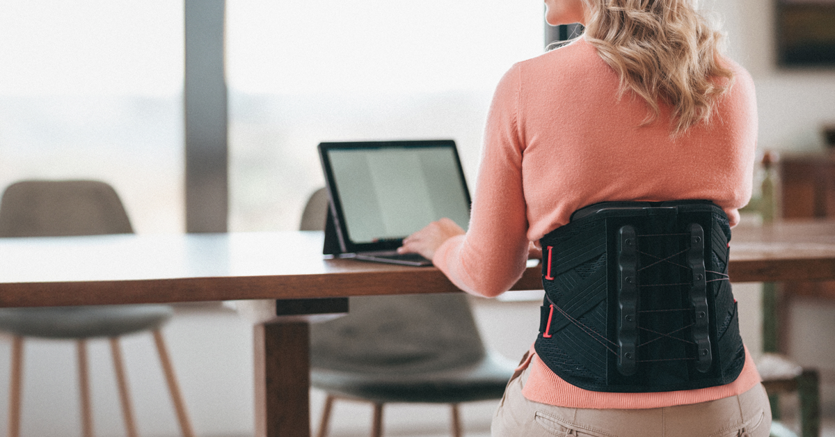
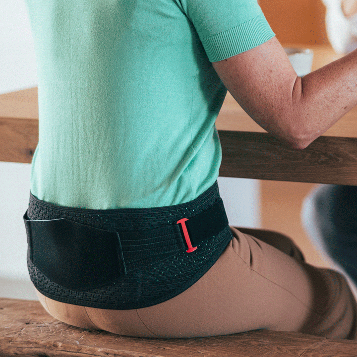
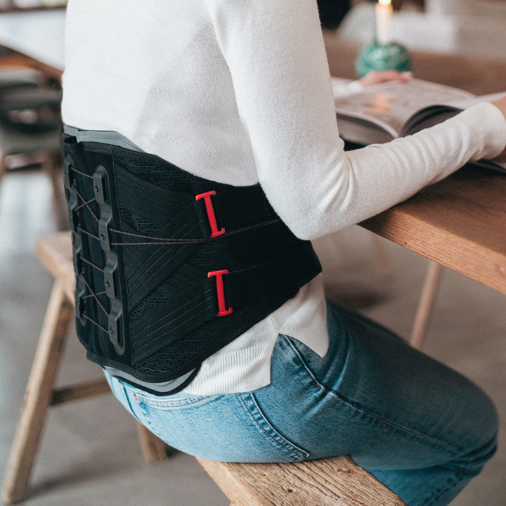
Low back pain is an increasingly widespread issue1 that can often be relieved through the use of a back brace2,3,4. The new DonJoy LumboForce® range provides a versatile option for low back pain sufferers, offering six distinct products that cater to various levels of lumbar spine support. From light support for mild discomfort to more advanced braces for severe conditions, this refreshed range of products embodies a modular approach to back care, ensuring both patients and healthcare professionals have access to the right tool for the job.
DonJoy LumboForce® Sacro: new targeted support for sacroiliac and pelvic stability
Specifically designed for stabilizing the pelvis and relieving pain in the sacroiliac (SI) joint, the new LumboForce Sacro features dual repositionable proprioception pads. These pads not only massage the affected area to promote blood circulation, but also offer flexible support tailored to the patient’s needs.
The brace’s semi-elastic webbing and bilateral straps ensure that compression is both supportive and adjustable, making it an excellent choice for conditions like symphysis insufficiency and sacroiliac joint trauma.
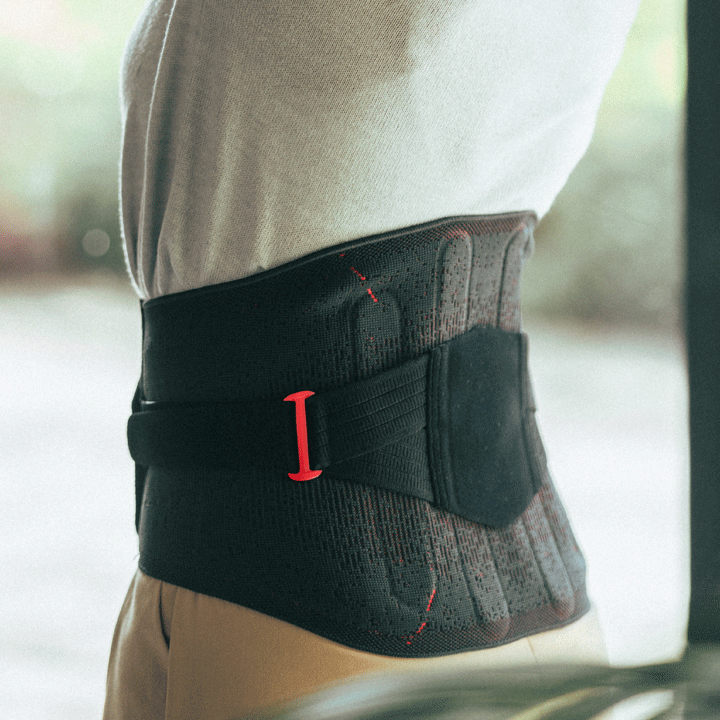
DonJoy LumboForce® 1: moderate support with comfort in focus
For patients needing moderate lumbar support, LumboForce 1 offers a balance of comfort and stability. It features flexible dorsal stays that support the affected spinal segments, providing support and helping to reduce pain.
The addition of a repositionable proprioception pad massages the lower back to help relieve discomfort. This brace is particularly suitable for those dealing with lumbalgia, osteochondrosis, or spondylarthrosis, conditions that benefit from moderate stabilization without restricting movement.
DonJoy LumboForce® 2: enhanced compression with customizable support
Taking the support up a level, the LumboForce 2 introduces bilateral straps that allow for greater control over compression. This feature is useful for patients who need variable support throughout the day, as it enables them to adjust the brace to their comfort.
Like LumboForce 1, this model includes flexible dorsal stays and a repositionable proprioception pad, making it suitable for managing both acute and chronic low back pain.
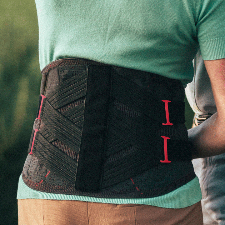
DonJoy LumboForce® 3: partial immobilization for severe lumbar conditions
LumboForce 3 is designed for patients requiring more substantial support and partial immobilization of the lumbar spine. This model can be particularly beneficial for conditions like spondylolisthesis, facet syndrome, and herniated discs in the early stages.
The adjustable dorsal stays and double bilateral straps provide a superior level of compression, while the option to choose between two heights (26 cm and 32 cm) helps ensure a proper anatomical fit for both men and women.
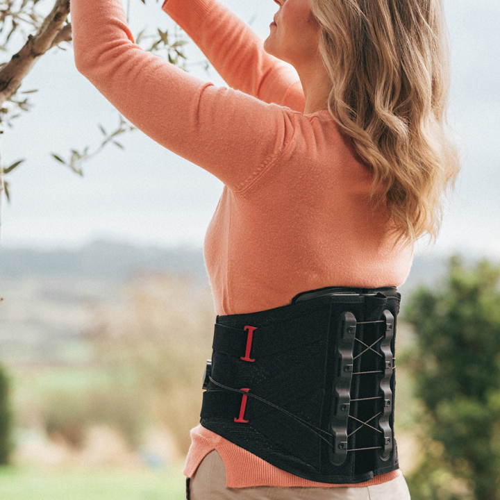
DonJoy LumboForce® 4: advanced stabilization for serious spinal issues
For those with more severe spinal conditions, such as advanced herniated discs or lumbar spinal canal stenosis, LumboForce 4 offers advanced support. This brace features a rigid aluminum back frame that bridges the lumbar spine, providing partial immobilization crucial for healing.
The bilateral pulley system and flexible dorsal stays work together to offer a higher level of compression and support, while the adjustable swiveling wings ensure the brace adapts to the patient’s movements. This design not only enhances comfort but also helps ensure that the brace remains effective throughout the healing process.
DonJoy LumboForce® 5: maximum immobilization for critical support
LumboForce 5 provides the highest level of support and immobilization in the range. This model is engineered for conditions that require greater lumbar immobilization, such as advanced stages of herniated discs, stable vertebral fractures, and severe spinal stenosis.
The rigid back shell, combined with dynamic side wings and a rigid abdominal pad, ensures over 50% circumferential waist support. The bilateral pulley system allows for precise compression adjustment, making this brace the go-to option for the most critical lumbar conditions.
Do back braces weaken abdominal muscles?
A common misconception about back braces is that wearing them can weaken your abdominal muscles. A 2019 study by Azadinia et al. showed that this is a false assumption, and that wearing a back brace for an average of more than 7 hours a day for 4 weeks did not lead to atrophy of deep trunk muscles, including abdominal muscles5.
Why Choose DonJoy LumboForce?
The DonJoy LumboForce range stands out not only for its comprehensive coverage of lumbar spine support needs but also for its focus on patient comfort and compliance. The range’s unisex design, anatomically shaped for an ergonomic fit, and use of soft, breathable materials help ensure that patients can wear these braces comfortably throughout the day. Whether you’re a healthcare provider seeking reliable options for your patients or someone dealing with back pain, the LumboForce range offers a well-rounded, customizable approach to lumbar care.
For more information on these products, visit our website.
References
Hoy, D., Bain, C., Williams, G., et al. (2012). A systematic review of the global prevalence of low back pain. Arthritis Rheum., 64(6): 2028-2037.
Mi, J., Ye, J., Zhao, X., Zhao, J. (2018). Effects of lumbosacral orthoses on postural control in individuals with or without non-specific low back pain. Eur Spine J., 27(1): 180-186.
Soo Choi, J., Kim, H., Lim, J., Seok Ryu, J. (2022). The facilitation of trunk muscles by abdominal bracing during walking in chronic low back pain patients. J Biomech, 143: 111299.
Ludvig, D., Preuss, R., Larivière, C. (2019). The effect of extensible and non-extensible lumbar belts on trunk muscle activity and lumbar stiffness in subjects with and without low-back pain. Clin Biomech (Bristol, Avon), 67: 45-51.
Azadinia, F., Ebrahimi Takamjani, I., Kamyab, M., Kalbassi, G., Sarrafzadeh, J., & Parnianpour, M. (2019). The Effect of Lumbosacral Orthosis on the Thickness of Deep Trunk Muscles Using Ultrasound Imaging: A Randomized Controlled Trial in Patients With Chronic Low Back Pain. American journal of physical medicine & rehabilitation, 98(7), 536–544.
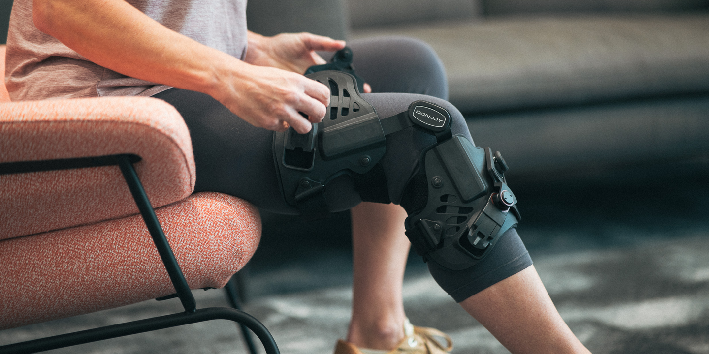
Living with knee osteoarthritis (OA) can significantly impact daily life, making even simple activities challenging. DonJoy OA GO® is a groundbreaking knee brace designed to help alleviate pain and improve mobility for those suffering from this condition. This innovative brace features several key components that work together to help provide effective relief and support.
Innovative design and functionality
The standout feature of the DonJoy OA GO knee brace is its unique three-point offloading system, which specifically targets and offloads the affected knee compartment. This system helps to reduce the pressure on the damaged area, thereby helping to alleviate pain and discomfort.
The brace also includes dual BOA® dials that allow for easy adjustments. With a simple turn of these dials, users can customize the level of support they need, ensuring a personalized fit that caters to their specific condition.
Comfort and stability
Comfort is crucial when it comes to wearing a knee brace for extended periods. The OA GO knee brace addresses this with flexible shells and anti-slip bands. These features help ensure that the brace stays in place and does not migrate during use, providing consistent support throughout the day.
Additionally, the brace is made from breathable, anti-bacterial material, which promotes hygiene and makes it suitable for long-term wear without causing irritation or discomfort.
Enhancing quality of life
The goal of the DonJoy OA GO knee brace is to help users maintain an active and fulfilling lifestyle despite their osteoarthritis. Whether you’re engaging in outdoor activities, climbing stairs, or playing with your grandchildren, this brace offers the support you need to help stay active and pain-free. The ability to adjust the brace easily means that you can tailor the support to your activity level, providing more stability or flexibility when you need it.
Versatility and availability
Half wrap, half sleeve, the OA GO knee brace combines the benefits of both to make fitting quick and easy. Simply open the thigh section then pull the lower sleeve section over the calf before securing the support with the hook-and-loop fastening.
Available in various sizes, the brace is a versatile option for individuals with different needs and body types, helping to ensure that patients can find a brace that fits them well and provides the necessary support. The ease of use and adjustability make it a convenient choice for managing mild to moderate knee osteoarthritis.
Living with knee osteoarthritis doesn’t have to mean giving up the activities you love. The DonJoy OA GO knee brace offers a practical and effective solution to manage pain and improve mobility. With its innovative design, adjustable support, and focus on comfort, this brace can help you lead a more active and pain-free life.
Living with knee osteoarthritis (OA) can be a challenging experience, marked by pain and limited mobility. For those navigating this condition, finding ways to manage symptoms and maintain an active lifestyle is crucial. This is where DonJoy® OA braces come into play, offering a range of innovative products designed to alleviate pain and support knee health.
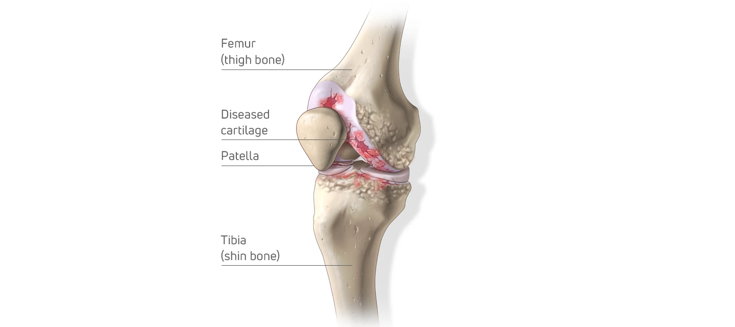
What is knee osteoarthritis?
Knee osteoarthritis (OA) is a degenerative joint disease characterized by the breakdown of cartilage in the knee joint. This deterioration leads to pain, stiffness, and swelling, making everyday activities challenging. Common risk factors include aging, obesity, joint injuries, and repetitive stress on the knee joints.
The role of bracing in knee OA management
Bracing is a non-invasive treatment option that can significantly reduce pain and improve function for individuals with knee OA.1 By redistributing weight away from the affected compartment of the knee, braces help decrease pressure on the affected areas, helping to provide relief and enhance mobility.2,3
Go Roam with DonJoy OA braces
DonJoy has established itself as a leader in the field of orthopedic bracing, offering a variety of braces designed for the full spectrum of knee OA severities. Two new products in their lineup include the DonJoy OA GO® and the ROAM™ OA.
DonJoy OA GO®: This brace is designed with a unique fitting mechanism that allows for easy adjustments, helping to provide personalized pain relief and support. It’s an excellent option for individuals looking for a customizable and comfortable brace to address mild to moderate single compartment osteoarthritis.
ROAM™ OA: As the latest innovation in offloader braces, the ROAM OA offers advanced features aimed at maximizing comfort and mobility. This brace is for active individuals who need robust support without compromising their movement. Designed for moderate to severe unicompartmental knee osteoarthritis.
Why choose DonJoy OA braces?
Innovation and quality: With a legacy of over forty years of knee bracing, DonJoy is renowned for its commitment to quality and continuous innovation, ensuring our braces meet the highest standards.
Patient-centered design: Our knee osteoarthritis braces are designed with the patient’s needs in mind, offering features that enhance comfort and ease of use.
Proven effectiveness: Clinical studies and patient testimonials highlight the effectiveness of DonJoy OA braces in reducing pain and improving quality of life.3,4,5
If you or someone you know is struggling with knee osteoarthritis, consider speaking to a healthcare professional about the braces offered by DonJoy. With the right brace, managing knee OA can become a more manageable part of daily life, allowing for continued activity and improved well-being.
For more information on knee OA and DonJoy OA braces, visit our official website: www.donjoyoabraces.com.
References
Brophy RH, Fillingham YA. AAOS Clinical Practice Guideline Summary: Management of Osteoarthritis of the Knee (Nonarthroplasty), Third Edition. J Am Acad Orthop Surg. 2022 May 1;30(9):e721-e729.
Nagai K, Yang S, Fu FH, Anderst W. Unloader knee brace increases medial compartment joint space during gait in knee osteoarthritis patients. Knee Surg Sports Traumatol Arthrosc. 2019 Jul;27(7):2354-2360.
Dries T, Van Der Windt JW, Akkerman W, Kluijtmans M, Janssen RPA. Effects Of A Semi-rigid Knee Brace On Mobility And Pain In People With Knee Osteoarthritis. J Rehabil Med Clin Commun. 2022 Jul 5;5:2483.
Khan SJ, Khan SS, Usman J, Mokhtar AH, Abu Osman NA. Orthoses versus gait retraining: Immediate response in improving physical performance measures in healthy and medial knee osteoarthritic adults. Proc Inst Mech Eng H. 2020 Jul;234(7):749-757.
Nagai K, Yang S, Fu FH, Anderst W. Unloader knee brace increases medial compartment joint space during gait in knee osteoarthritis patients. Knee Surg Sports Traumatol Arthrosc. 2019 Jul;27(7):2354-2360.
Anterior cruciate ligament (ACL) injuries are a common concern in sports and physical activities, often requiring extensive rehabilitation and sometimes even surgery. Knee braces are one of the most prescribed devices in the orthotic industry, with medical device companies such as Enovis™ supplying a range of knee bracing solutions for ACL protection and injury prevention. But despite their widespread use, the question remains: Do ACL braces really work?
This article explores the world of ACL braces, examining their purported benefits and the scientific evidence behind their effectiveness, before presenting a new product for people looking to safeguard their knee health.
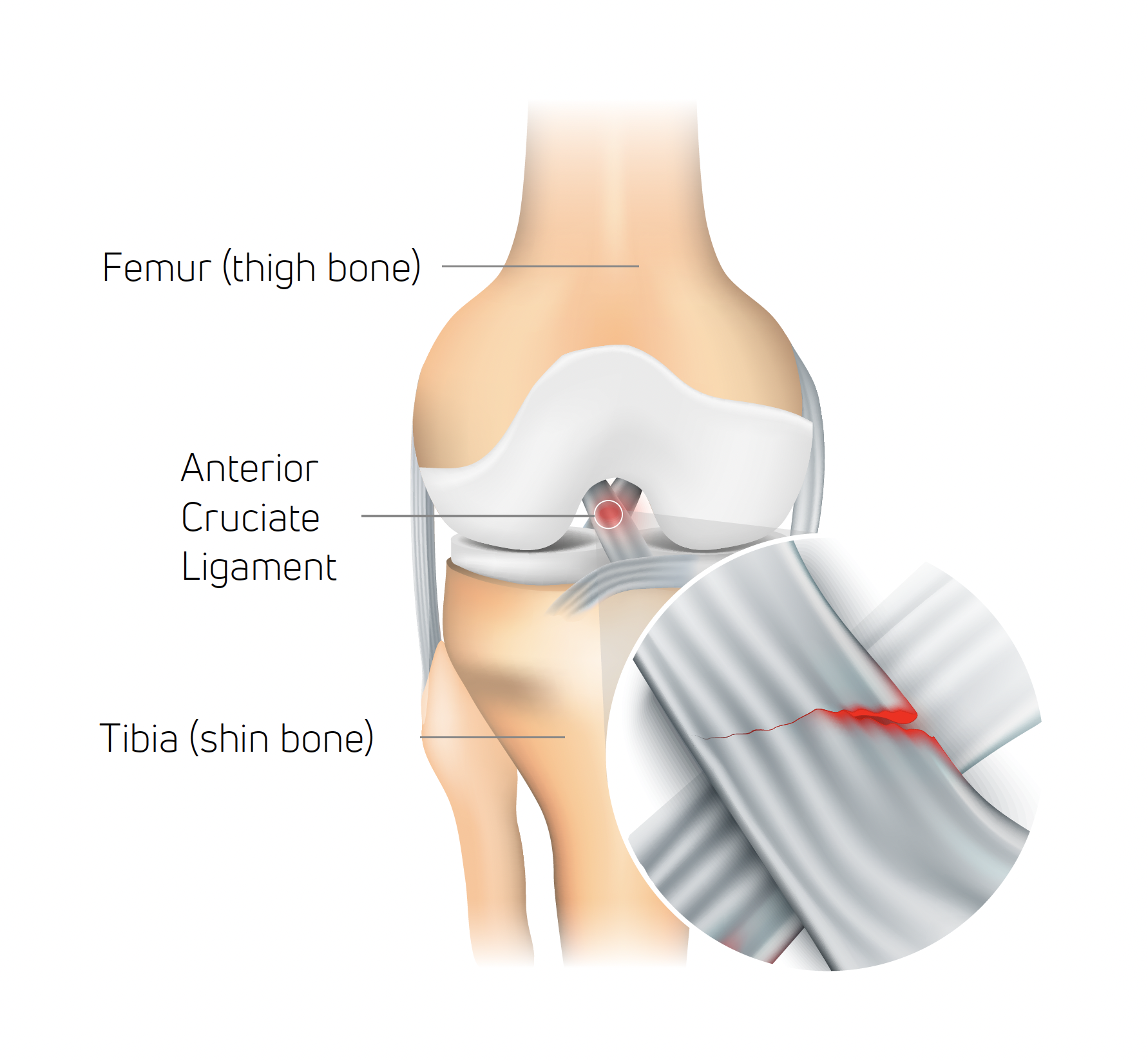
ACL injury: definition and causes
The anterior cruciate ligament (ACL) serves as an essential stabilizer within the knee joint, preventing the tibia (shine bone) from shifting forward in relation to the femur (thigh bone) and ensuring rotational stability.
While an ACL tear can occur due to excessive external force applied to the knee, it’s distinctive in that it can also happen without direct contact, which accounts for 70% of reported ACL injuries1.
In sports like football and other field/court activities, non-contact ACL injuries usually occur during abrupt stops, sudden changes in direction, or when landing from a jump with insufficient knee and hip flexion (at or near full extension)2. The typical scenario involves a combination of deceleration, directional change while the foot is planted, and the knee being near or fully extended. This action can put excessive twisting force on the ACL, leading to strain or rupture.
Evidence that wearing a knee brace can help prevent ACL injury
Clinical studies have demonstrated that wearing a knee brace during activity can help prevent ACL injury as well as protect against reinjury3,4,5.
In a systematic review of current evidence carried out in 2023, Tuang et al. found that protective knee braces were able to control forwards and backwards and sideways knee motion and decrease ACL load/strain during high-risk maneuvers, which may in turn decrease the risk for non-contact ACL injuries3.
With around half of ACL injuries occurring in 15–25-year-olds6, knee bracing effectiveness for young people is a key concern for many. Perrone et al.’s 2019 study involved prescribing knee braces to a group of adolescents post-ACL surgery. The results showed that post-operative use of functional bracing can result in reduced reinjury following ACL reconstruction4.
Bodendorfer et al.’s 2013 study also recommended knee bracing for ACL patients. It found that people with ACL-deficient knees can benefit from the control and proprioception functional bracing can offer. And for highly active athletes participating in high-impact sports, knee bracing further offers protection to the knee ligaments and meniscus during impact from the side5.
How DonJoy® knee braces help prevent ACL injury
DonJoy® is a name synonymous with knee bracing. A key brand of Enovis, it has been manufacturing and supplying braces for knee ligament protection since the late 1970s, using patented technology that reduces ACL strain.
The Four-Points-of-Leverage™ system featured in DonJoy knee braces consists of a rigid cuff and strap configuration. Through this, a posterior force is applied to the tibia, which prevents anterior movement and reduces the strain on the ACL7.
The second key technology in DonJoy knee braces is the FourcePoint® hinge. This complements the Four-Points-of-Leverage design by damping knee joint extension, which improves the mechanical performance of the brace and reduces shear forces at the knee. The hinge resistance kicks in during the last 25 degrees of knee extension, targeting the vulnerable “at-risk” position.
When combined, the FourcePoint hinge and the Four-Points-of-Leverage design create a more comfortable brace that diminishes anterior shear forces on the knee. This stability is particularly advantageous for people wanting to prevent ACL injuries during activity and those recovering from ACL injuries, as it eases strain on the deficient or healing ACL graft8,9.
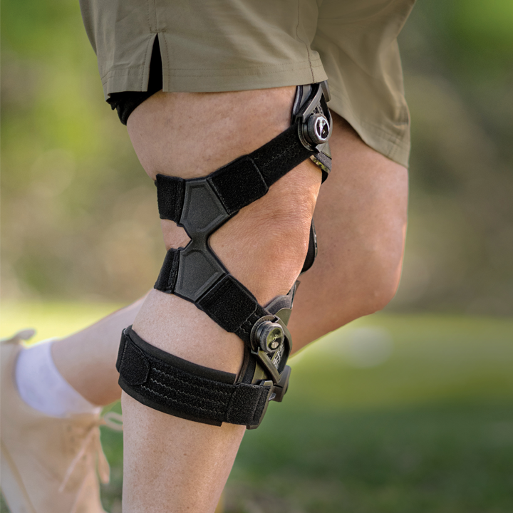
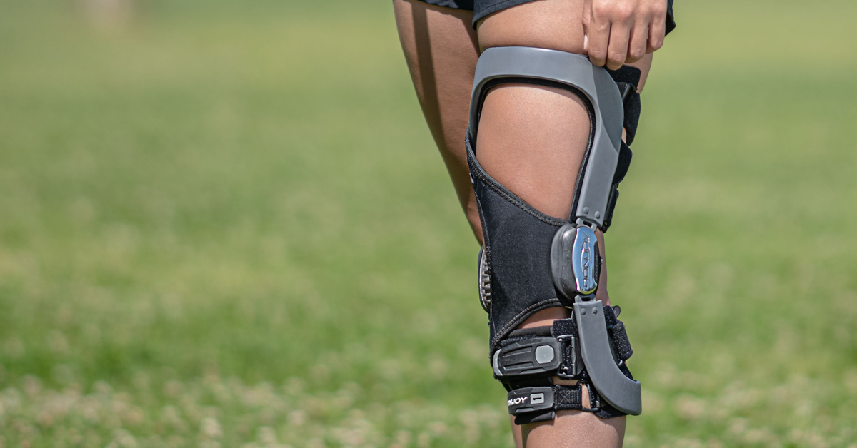
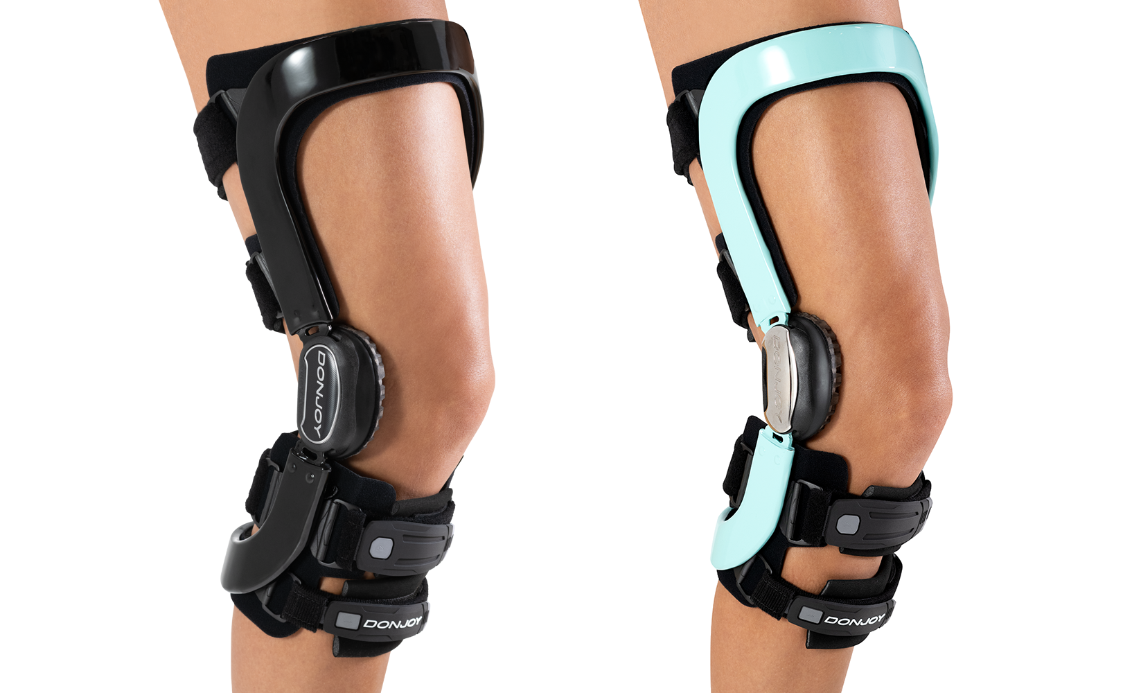
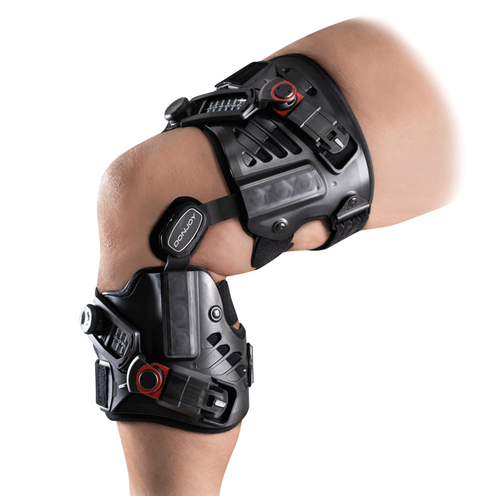
Defiance® PRO: custom knee ligament bracing from DonJoy
When it comes to ligament knee bracing, few product names stand out more than Defiance®. Alongside off-the-shelf alternatives, DonJoy’s flagship custom brace has been protecting knees for decades. Now with the Defiance® PRO taking the design to the next level, those looking to prevent ACL injuries have a new name to trust in.
Featuring the proven Four-Points-of-Leverage and FourcePoint technologies, Defiance PRO also provides a range of customizable elements to offer patients an enhanced wearing experience.
Every Defiance PRO order begins with the patient receiving a precise measurement of their leg from an Enovis representative. These measurements are then used to build a brace exactly matched to the customer’s leg for an even closer and more comfortable fit.
DonJoy Defiance PRO knee ligament brace
Patients can further tailor their brace by choosing the frame colour from over 30 available options and adding a series of accessories, including a sports cover and silicone condyle pads for extra comfort.
With this combination of clinically proven technology and superior craftmanship, patients can be confident that DonJoy is the name to trust for knee ligament bracing.
Boden BP, Dean GS, Feagin JA Jr, Garrett WE Jr. Mechanisms of anterior cruciate ligament injury. Orthopedics. 2000 Jun;23(6):573-8.
Silvers, H. J., & Mandelbaum, B. R. (2007). Prevention of anterior cruciate ligament injury in the female athlete. British journal of sports medicine, 41 Suppl 1(Suppl 1), i52–i59.
Tuang, B.H.H., Ng, Z.Q., Li, J.Z., Sirisena D. (2023). Biomechanical Effects of Prophylactic Knee Bracing on Anterior Cruciate Ligament Injury Risk: A Systematic Review. Clin J Sport Med. Jan 1;33(1):78-89.
Perrone, G.S., Webster, K.E., Imbriaco, C., Portilla, G.M., Vairagade, A., Murray, M.M., Kiapour, A.M. (2019). Risk of Secondary ACL Injury in Adolescents Prescribed Functional Bracing After ACL Reconstruction. Orthop J Sports Med. Nov 12;7(11):2325967119879880.
Bodendorfer, B.M., Anoushiravani, A.A., Feeley, B.T., Gallo, R.A. (2013). Anterior cruciate ligament bracing: evidence in providing stability and preventing injury or graft re-rupture. Phys Sportsmed. Sep;41(3):92-102.
Griffin LY, Albohm MJ, Arendt, EA, et al. (2006). Understanding and Preventing Noncontact Anterior Cruciate Ligament Injuries: A Review of the Hunt Valley II Meeting, January 2005. Am J Sports Med 34(9):1512-32
Fleming, B. C., Renstrom, P. A., Beynnon, B. D., Engstrom, B., & Peura, G. (2000). The influence of functional knee bracing on the anterior cruciate ligament strain biomechanics in weightbearing and nonweightbearing knees. The American journal of sports medicine, 28(6), 815–824.
Théoret, D., & Lamontagne, M. (2006). Study on three-dimensional kinematics and electromyography of ACL deficient knee participants wearing a functional knee brace during running. Knee surgery, sports traumatology, arthroscopy : official journal of the ESSKA, 14(6), 555–563.
Stanley, C. J., Creighton, R. A., Gross, M. T., Garrett, W. E., & Yu, B. (2011). Effects of a knee extension constraint brace on lower extremity movements after ACL reconstruction. Clinical orthopaedics and related research, 469(6), 1774–1780.
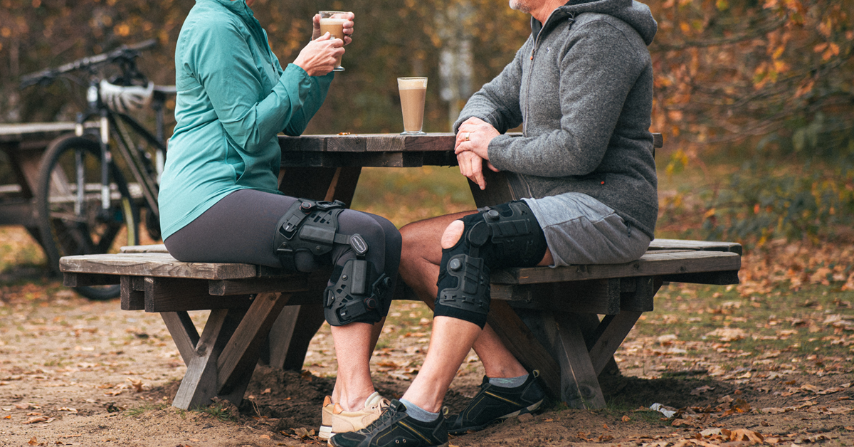
Knee osteoarthritis (OA) is a painful condition that affects over 650 million people worldwide1. Occurring predominantly in those aged 40 and over, the barrier to activity it presents can have a debilitating effect on both the physical and mental health of sufferers. Thankfully, knee braces have been shown to have a positive effect on the symptoms of OA2, and with the release of ROAM™ OA, DonJoy® has a new way to help OA patients reduce knee pain and stay active and healthy.
Comfortable, easy to use, and effective
Regular use of offloader knee braces has been shown to contribute to improved daily functioning, reduced pain, and enhanced mobility, ultimately leading to a better quality of life2. ROAM OA is designed to improve mobility and provide relief by unloading the pressure of moderate to severe unicompartmental osteoarthritis or other knee pain. Its lightweight, low-profile joint offloading and support system offer a high level of comfort while remaining user-friendly for both prescribers and patients.
Designed for diverse patient needs
Whether for conservative care or as a preparation for surgery, ROAM OA’s series of adjustable settings cater to a wide range of patients looking to enhance their activity levels and maintain an active lifestyle.
To address the most challenging fitting needs, ROAM OA is also available in a custom-made version. Tailored to individual measurements, this option ensures a unique fit with custom-positioned condyle and uprights, optimal height setting, and personalized cuff sizes and strap lengths.
Features and benefits
With a patient-focused approach, ROAM OA’s list of features ensures ease of use and aids compliance.
Once set by a trained fitter, the “set and forget” straps provide a visual guide for patients to help make donning and removing the brace simple and worry-free.
The patented Mag-Align magnetic buckles provide an audible “snap” on connection, instilling confidence in patients, even those with eyesight or dexterity challenges.
The BOA® Fit System allows patients to easily dial in support and pain relief on demand. Simply turn the dials to increase tension in the brace, or pull them out for quick release.
And patent-pending comfort straps and a condyle harness help negate migration of the brace while enhancing comfort by leaving the popliteal fossa area behind the knee free from friction.
*BOA® is a registered trademark of BOA® Technology Inc.
References
Cui A, Li H, Wang D, Zhong J, Chen Y, Lu H. Global, regional prevalence, incidence and risk factors of knee osteoarthritis in population-based studies. EClinicalMedicine. 2020 Nov 26;29-30:100587.
Feehan NL, Trexler GS, Barringer WJ. The Effectiveness of Off-Loading Knee Orthoses in the Reduction of Pain in Medial Compartment Knee Osteoarthritis: A Systematic Review. J Prosthet Orthot 2012;24(1):39-49.
As winter sports enthusiasts hit the slopes, the importance of protecting vulnerable joints cannot be overstated. For skiers and snowboarders, the rigors of downhill adventures can take a toll on the knees. Thankfully DonJoy® knee braces are clinically proven to protect the knee and safeguard against ligament injuries.1 Here are ten compelling reasons why skiers and snowboarders should consider wearing a DonJoy rigid knee ligament brace.
1. Dynamic Stability
The unpredictable terrain of snowy slopes demands dynamic stability. DonJoy knee braces feature the Four-Points-of-Leverage™ system, a combination of a rigid cuff and straps that provides a constant dynamic load and is clinically proven to reduce anterior cruciate ligament (ACL) strain.2
2. Prevent Ligament Injuries
The lateral movements and sudden stops inherent in skiing and snowboarding can put stress on knee ligaments, and when it comes to ligament injuries, prevention is better than any cure. DonJoy’s patented FourcePoint® Hinge keeps the knee out of the “at risk” position, helps prevent anterior tibial translation, and dampens knee joint extension, helping to protect the knee from injury.3
3. Post-Injury Protection
With a re-injury rate of 5-10% for people who have suffered an ACL injury, protecting the knee is essential.4 The Four-Points-of-Leverage technology featured on DonJoy knee braces effectively reduces ACL strain, which can be crucial during rehabilitation while the graft is remodelling.2
4. Enhance confidence
The protection offered by a DonJoy brace can give skiers and snowboarders the confidence to push their limits with greater assurance.
5. Important for women and young people
Of those who suffer ligament injuries, women and young people are the most susceptible. With 50% of ACL injuries occurring in 15-25 year olds,5 and women 8x more likely to injure an ACL,6 it is even more essential for them to wear a knee brace while skiing or snowboarding.
6. Unique braces made to measure
Every Defiance and A22® knee brace is made to the specific measurements of the customer’s leg for an even closer and more comfortable fit.
7. Off-the-shelf options for a fast fix
For customers not looking for a custom option, DonJoy offers a wide choice of off-the-shelf knee braces. Armor™ comes in 7 different sizes of thigh circumference for fast and simple measuring and ordering.
8. Customise your brace to suit
DonJoy’s Defiance knee braces offer a range of customisable features to suit customers. Along with optional accessories including a sports cover and silicone condyle pads, these braces are also available in over 30 frame colours and patterns.
Defiance knee braces
9. DonJoy braces won’t get in the way
Thanks to their lightweight, low-profile design, DonJoy knee braces fit easily under ski trousers and snowboard pants. Many of the braces are also available in a short calf length to ensure boot clearance.
10. There’s more than just bracing
DonJoy knee braces are complemented by a huge range of medical devices provided by its co-brands in the Enovis™ organisation. Whether it is cold therapy products to reduce pain following injury, or electro muscle stimulation devices to aid in recovery, Enovis offers support for skiers and snowboarders throughout the continuum of care.
Ackerman DR et al. Prophylactic Knee Bracing in Offensive Linemen of the National Football League: A Retrospective Analysis of Usage Trends, Player Performance, and Major Knee Injury. Orthop J Sports Med. 2023 Aug 25;11(8):23259671231191767.
Fleming BC et al. The influence of functional knee bracing on the anterior cruciate ligament strain biomechanics in weightbearing and nonweightbearing knees. Am J Sports Med 2000;28(6):815-24.
Yu B et al. Immediate effects of a knee brace with a constraint to knee extension on knee kinematics and ground reaction forces in a stop-jump task. Am J Sports Med 2004;32(5):1136-43.
Arendt EA et al. Anterior cruciate ligament injury patterns among collegiate men and women. Journal of Athletic Training. 1999;34(2):86-92.
Griffin LY et al. Understanding and Preventing Noncontact Anterior Cruciate Ligament Injuries: A Review of the Hunt Valley II Meeting, January 2005. American Journal of Sports Medicine. 2006 34:9. 1512-1532.
Mancino F et al. Anterior cruciate ligament injuries in female athletes. Bone Joint J. 2023 Oct 1;105-B(10):1033-1037.
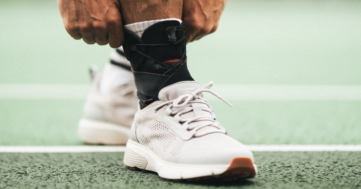
Lateral ankle sprains are one of the most common sports-related injuries1; in the United States alone, more than 23,000 people suffer a lateral ankle sprain every day.2 To help address this situation, DonJoy® introduces the new DonJoy ActyLight®, an ankle support so comfortable and easy to fit, you’ll forget you’re even wearing it.
What is an ankle sprain, and why do they happen?
When the ligaments of the ankle are damaged, this is called a sprain. If the foot suddenly rolls inward during activity, the subsequent forceful ankle plantarflexion and inversion can result in stretching and tearing of the ankle ligament fibers. As well as causing swelling and bruising, sprains are usually painful, especially when the person attempts to put weight on the foot.
Ankle sprains are categorized into three grades of severity:
Grade I (Mild): This involves minor stretching and tiny tears in the ligament fibers, leading to mild tenderness and swelling around the ankle.
Grade II (Moderate): In this grade, there is partial tearing of the ligament, resulting in moderate tenderness and swelling around the ankle. Certain movements can cause some abnormal looseness in the ankle joint.
Grade III (Severe): This is the most severe grade, where the ligament is completely torn. It leads to significant tenderness and swelling around the ankle, and certain movements can cause substantial instability in the ankle joint.
What is chronic ankle instability?
Approximately 40% of individuals who suffer an ankle sprain later develop chronic ankle instability (CAI) and report persistent symptoms.3,4 This condition is marked by sensations or instances of the ankle unexpectedly giving way.5
People with CAI commonly experience ongoing symptoms such as persistent swelling, pain, weakness, restricted ankle movement, instability, reduced self-reported functionality, and recurrent ankle sprains.5,6
CAI has been recognized as a precursor to ankle osteoarthritis (OA), with its onset typically happening a decade earlier than knee or hip OA.7
Chronic ankle sprains might necessitate surgical intervention through arthroscopic ligament reconstruction. Individuals with CAI are noted to exhibit both mechanical instability, related to structural changes around the ankle, and functional instability, which is associated with decreased sensorimotor and neuromuscular control.8
Bracing for chronic ankle instability
An ankle brace is worn to support and stabilize the ankle, either as a preventive measure or after an injury has occurred. These braces come in soft or semi-rigid varieties and are intended for one or more of the following purposes:
Improve ankle stiffness and thus mechanical stability9
Improve neuromuscular control10
Improve grounding of the foot10
Decrease excessive range of motion (ROM)9
Enhance proprioceptive acuity (the body’s ability to sense its own location, movement, and actions)11
Numerous clinical studies have been conducted to assess the effectiveness of ankle bracing in these areas.
In 1998, Vaes et al. discovered that the Aircast® Air-Stirrup® brace significantly reduced talar tilt in unstable ankles during static and dynamic tests, and it slowed down the simulated sprain speed.12
In a study from 2000, Hals et al. demonstrated a significant enhancement in shuttle-run performance among subjects with post-acute, mechanically stable yet functionally unstable ankle sprains when using the Aircast Sport Stirrup® brace.13
A randomized controlled trial conducted by Janssen et al. in 2014, involving 384 athletes who had experienced a lateral ankle sprain, revealed that using an Aircast A60™ brace was more effective than neuromuscular training in reducing the recurrence of ankle sprains.14
Habadi et al. (2014) showed the advantages of soft and semi-rigid ankle orthoses in improving the dynamic balance of individuals with functional ankle instability.15
And a 2020 systematic review by Reyburn and Powden concluded that the current body of research strongly supports the positive impact of ankle braces on the dynamic balance of individuals with CAI.16
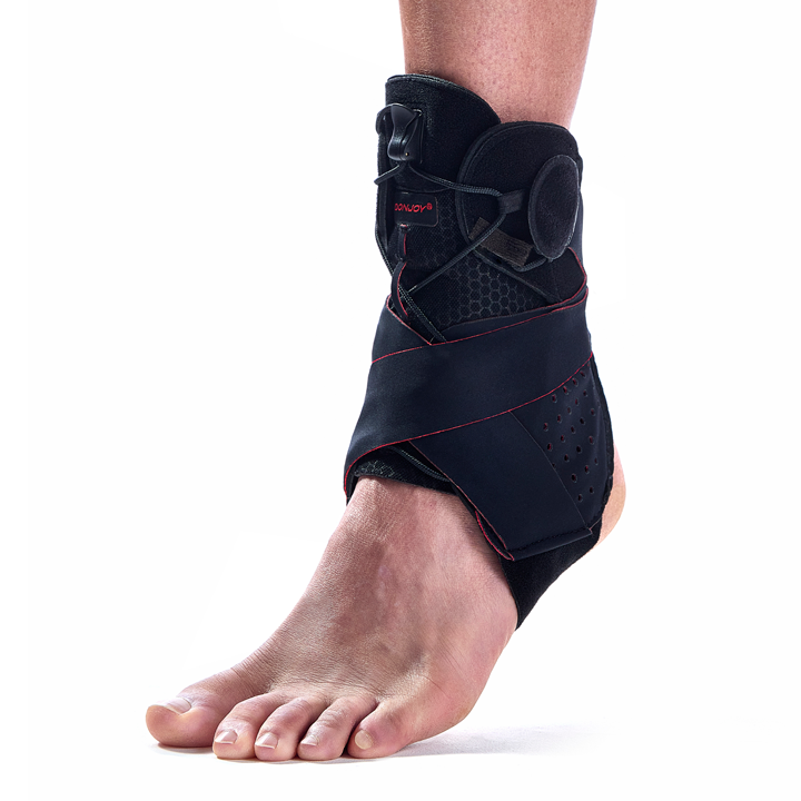
Introducing ActyLight by DonJoy®: fit-and-forget ankle support
The convenience and comfort of the DonJoy ActyLight® ankle support means patients will forget they’re even wearing it.
Designed to deliver stability and protection for mild to moderate lateral ankle sprains, thanks to its removable bilateral stays, quick lace-locking mechanism, and step-in design, this modular brace can also be used for the prevention of ankle injuries.
All of this means that patients can rely on ActyLight throughout their journey of activity, from healthy, active use, to support following injury, and to prevention of reinjury in the future.
Fong, D.T.P., Hong, Y., Chan, L.K., Yung, P.S.H. and Chan, K.M., (2007). A systematic review on ankle injury and ankle sprain in sports. Sports medicine, 37(1), pp.73-94.
Hubbard, T.J. and Wikstrom, E.A., (2010). Ankle sprain: pathophysiology, predisposing factors, and management strategies. Open Access Journal of Sports Medicine, 1, p.115.
Anandacoomarasamy, A. & Barnsley, L. (2005). Long term outcomes of inversion ankle injuries. Br J Sports Med, 39(3): e14; discussion e14.
Konradsen L., Bech L., Ehrenbjerg M. & Nickelsen T. (2002). Seven years follow-up after ankle inversion trauma. Scand J Med Sci Sports, 12(3): 129-135.
Hertel, J. & Corbett, R.O. (2019). An updated model of chronic ankle instability. Journal of athletic training, 54(6): 572-588.
Ahn, C. S., Kim, H. S., & Kim, M. C. (2011). The Effect of the EMG Activity of the Lower Leg with Dynamic Balance of the Recreational Athletes. The Journal of Physical Therapy Science. 579–583.
Saltzman, C.L., Zimmerman, M.B., O’Rourke, M., Brown, T.D., Buckwalter, J.A. & Johnston, R. (2006). Impact of comorbidities on the measurement of health in patients with ankle osteoarthritis. J Bone Joint Surg Am., 88(11): 2366-2372.
Hertel, J., (2002). Functional anatomy, pathomechanics, and pathophysiology of lateral ankle instability. Journal of athletic training, 37(4): 364.
Zinder, S.M., Granata, K.P., Shultz, S.J. & Gansneder, B.M. (2009). Ankle bracing and the neuromuscular factors influencing joint stiffness. Journal of Athletic Training, 44(4): 363-369.
Kobayashi, T., Saka, M., Suzuki, E., Yamazaki, N., Suzukawa, M., Akaike, A., Shimizu, K. & Gamada, K. (2014). The effects of a semi-rigid brace or taping on talocrural and subtalar kinematics in chronic ankle instability. Foot & Ankle Specialist, 7(6): 471-477.
Raymond, J., Nicholson, L.L., Hiller, C.E. & Refshauge, K.M. (2012). The effect of ankle taping or bracing on proprioception in functional ankle instability: a systematic review and meta-analysis. Journal of Science and Medicine in Sport, 15(5): 386-392.
Vaes, P. H., Duquet, W., Casteleyn, P. P., Handelberg, F., & Opdecam, P. (1998). Static and dynamic roentgenographic analysis of ankle stability in braced and nonbraced stable and functionally unstable ankles. The American journal of sports medicine, 26(5): 692–702.
Hals, T. M., Sitler, M. R., & Mattacola, C. G. (2000). Effect of a semi-rigid ankle stabilizer on performance in persons with functional ankle instability. The Journal of orthopaedic and sports physical therapy, 30(9), 552–556.
Janssen, K. W., van Mechelen, W., & Verhagen, E. A. (2014). Bracing superior to neuromuscular training for the prevention of self-reported recurrent ankle sprains: a three-arm randomised controlled trial. British journal of sports medicine, 48(16): 1235–1239.
Hadadi, M., Mousavi, M. E., Fardipour, S., Vameghi, R., & Mazaheri, M. (2014). Effect of soft and semirigid ankle orthoses on Star Excursion Balance Test performance in patients with functional ankle instability. Journal of science and medicine in sport, 17(4): 430–433.
Reyburn, R. J., & Powden, C. J. (2020). Dynamic Balance Measures in Healthy and Chronic Ankle Instability Participants While Wearing Ankle Braces: Systematic Review With Meta-Analysis. Journal of sport rehabilitation, 30(4): 660–667.